Advanced Parathyroid Scan (Sestamibi Scintigraphy)
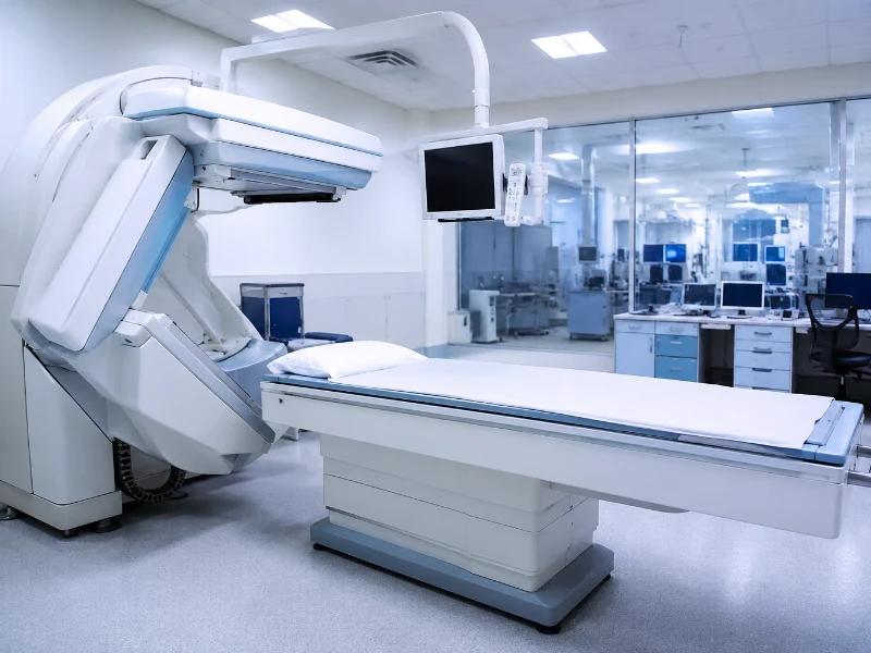
A Parathyroid Scan, clinically known as a Technetium (99mTc) Sestamibi scan, is a highly specialized endocrine nuclear medicine imaging procedure designed to identify and locate overactive parathyroid glands. While most individuals have four tiny parathyroid glands situated directly behind the thyroid gland in the neck, these nodules regulate the body’s precise systemic calcium balances. Specifically, by utilizing advanced dual-phase gamma cameras or hybrid SPECT-CT scanners, our diagnostic facility traces cellular radiotracer uptake to pinpoint hyperfunctioning tissue, such as benign adenomas or diffuse cellular hyperplasia, with outstanding precision.
Furthermore, because chronic overproduction of parathyroid hormone (PTH) continuously strips calcium from your skeletal structures, it can lead to severe osteoporosis, recurring kidney stones, digestive changes, and chronic fatigue. Standard cross-sectional structural imaging like routine ultrasounds frequently fails to distinguish tiny parathyroid tissue from surrounding thyroid nodules, making functional nuclear tracking essential. Consequently, our dedicated protocols utilize specialized delayed washout tracking to isolate hyperactive cellular clusters. As a result, endocrinologists and endocrine surgeons receive a highly clear map to plan targeted, minimally invasive surgical removals.
What is a Parathyroid Sestamibi Scan?
A parathyroid scan is a functional localization imaging panel that relies on the unique mitochondrial tracking properties of the Sestamibi radiotracer. Therefore, precise multi-tiered laboratory technology allows our nuclear medicine physicians to accurately trace tracer washing phases, identify hyperactive locations, and differentiate abnormal parathyroid nodules from normal thyroid tissues for a comprehensive endocrine review.
- Early Phase Imaging (Evaluating baseline tracer distribution across thyroid and parathyroid tissues)
- Delayed Phase Tracking (Mapping target areas where the tracer clears from normal tissue but remains trapped in overactive nodules)
- SPECT-CT Hybrid Cross-References (Providing 3D anatomical and structural localization mapping)
- Serum Calcium and Intact PTH biochemical correlations
Parathyroid Scan Diagnostic Pricing Guide
To ensure complete financial transparency for our patients, we provide clear, upfront cost structures across our advanced endocrine imaging services. Below is the current price breakdown for parathyroid imaging variations and associated biochemical panel matchings at our diagnostic laboratory hubs:
| Diagnostic Scan / Panel Variation | Estimated Cost (INR) |
|---|---|
| Standard Dual-Phase Parathyroid Scan | ₹6,000 – ₹7,500 |
| Advanced SPECT-CT Parathyroid Scan | ₹9,000 – ₹11,500 |
| Comprehensive Hyperparathyroidism Evaluation Panel | ₹10,500 – ₹13,000 |
Note: Final pricing variations can occur based on specialized pharmaceutical requirements or specific clinical instructions provided by your referring consultant.
Why is a Parathyroid Scan Performed?
Physicians commonly recommend a parathyroid cholescintigraphy evaluation when blood tests show elevated calcium levels alongside high parathyroid hormone numbers, or when patients suffer from chronic symptoms of hypercalcemia. Moreover, the analytical findings directly assist endocrinologists, endocrine surgeons, nephrologists, and rheumatologists in establishing precise medical plans. Our experienced clinical team ensures you receive highly accurate imaging and reports.
- Investigating the root biological cause of Hypercalcemia (abnormally high blood calcium levels)
- Locating a solitary benign tumor (Parathyroid Adenoma) before scheduled surgical treatment
- Investigating recurring kidney stones (Nephrolithiasis) driven by high calcium filtering
- Evaluating severe, unexplained bone thinning (Osteoporosis) or frequent skeletal fractures
- Pinpointing ectopic parathyroid tissue that has formed abnormally in the deep chest or mediastinum cavity
- Differentiating multi-gland hyperplasia from a single overactive parathyroid nodule
- Re-evaluating persistent or recurring hyperparathyroidism after a previous neck operation
- Investigating vague signs of chronic calcium excess, like persistent bone pain, muscle weakness, and fatigue
Key Diagnostic Extraction Phases Tracked During the Scan
Early Radiotracer Distribution
Approximately 10 to 15 minutes after the tracer injection, our advanced gamma camera captures initial images of your neck. During this initial phase, the Sestamibi compound accumulates naturally within both normal thyroid tissue and overactive parathyroid cells.
Delayed Washout Separation
We take follow-up images 2 to 3 hours later. Because normal thyroid tissue flushes out the tracer quickly while overactive parathyroid adenomas retain it for much longer, this crucial delay phase shows hyperactive glands glowing brightly against a dark background.
SPECT-CT 3D Fusion Mapping
When requested, our hybrid system rotates around your neck to combine functional nuclear images with highly detailed structural CT scans. This 3D mapping tells the surgeon exactly how deep the gland sits relative to your airway and blood vessels.
Conditions Managed and Monitored via Endocrine Scintigraphy
An accurate parathyroid scan helps clinicians identify, track, and manage a variety of metabolic, skeletal, and mineral disorders. You can find detailed endocrine tracking standards from the Endocrine Society clinical standards.
- Primary Hyperparathyroidism (Driven by a single or double parathyroid adenoma)
- Secondary and Tertiary Hyperparathyroidism (Driven by chronic kidney filtering issues)
- Parathyroid Hyperplasia (Diffuse overactivity across all four glands)
- Ectopic Parathyroid Adenomas (Glands hidden in the chest cavity or carotid sheath)
- Hypercalcemia-induced Osteopenia and severe bone thinning tracking
- Recurrent or persistent post-surgical hyperparathyroidism surveillance
- Parathyroid cysts and rare tissue anomalies tracking
If your endocrine screening reveals complex mineral variations or hormone elevations, your physician may suggest matching your imaging with other advanced diagnostic evaluations. For a detailed biochemical look at your circulating calcium and thyroid hormones, check out our specialized Hormone Test services or our focused Thyroid Profile test services. For complete organ processing and filtration checks, see our Kidney Function Test services or our Liver Function Test services. You can also explore our general blood test services, verify immune baselines with a Complete Blood Count (CBC) test, monitor blood sugar levels via an HbA1c test, or see how structural neuroendocrine systems are monitored using our specialized MRI Pituitary Brain services. If you need other standard blood checkups completed safely without traveling, you can book a certified mobile expert through our professional home blood collection services.
Preparation for a Parathyroid Scan
- Fasting is typically not required for a standalone parathyroid scan; you can eat and drink normally before your appointment.
- Drink plenty of plain water before your scan to remain well-hydrated, which helps make placing the intravenous line smooth and simple.
- Inform our laboratory staff about all daily medications, prescriptions, or nutritional supplements you take.
- Let our team know if you take regular calcium pills, high-dose Vitamin D, or thyroid hormone medications, as certain prescriptions should be reviewed by your doctor before testing.
- Avoid undergoing any diagnostic procedures involving iodinated contrast media (like a contrast CT scan) for at least 2 to 4 weeks before your appointment, as iodine can interfere with tracer extraction.
What to Expect During the Imaging Process
- Our professional nuclear medicine team registers you and reviews your endocrine testing history in our imaging suite.
- A certified nurse places a small intravenous (IV) line into a vein in your arm or hand.
- A safe, targeted dose of the Sestamibi tracking agent is introduced smoothly through the IV line.
- You will lie comfortably flat on your back on a cushioned imaging table with your head tilted back slightly.
- The advanced gamma camera moves close to your neck and upper chest area to capture the initial 15-minute early phase images.
- You will be free to relax in our waiting area for 2 to 3 hours while the tracer washes through your tissues.
- You will return to the scanner table for the delayed phase and SPECT-CT tracking, capturing the final 3D location details.
The entire diagnostic process, including the resting washout period, requires approximately 3 to 4 hours to complete safely.
Frequently Asked Questions
What is the difference between a thyroid scan and a parathyroid scan?
A thyroid scan checks the function of the larger thyroid gland using iodine or technetium tracers to evaluate metabolism or nodules. A parathyroid scan uses a specialized Sestamibi tracer that is retained specifically by overactive parathyroid glands, helping doctors find these tiny nodules even when hidden behind or inside the thyroid.
Is the Sestamibi tracer safe, and are there any side effects?
Yes. The Sestamibi tracking compound is exceptionally safe and widely used. It uses a very low, diagnostic dose of radiation that leaves your body naturally through your urine within 24 hours. The injection does not cause drowsiness or nausea, meaning you can drive home and resume normal activities right after the test.
Why do I have to wait several hours between the first and second scan phases?
The waiting period is critical because it allows the tracer to naturally wash out of your normal thyroid tissues. Overactive parathyroid adenomas hold onto the tracer much longer, meaning the delay phase is what allows the hyperactive glands to stand out clearly on the final images.
How long does it take to get my verified parathyroid scan results?
Our nuclear medicine facility processes and fuses your planar and SPECT-CT datasets efficiently. A senior specialist reviews the localized uptake charts, and a verified interpretation report is usually ready for your doctor within 24 hours.
Why is a nuclear scan preferred over managing high calcium levels with diet?
Ultimately, while dietary changes are excellent for general wellness, high blood calcium driven by a parathyroid adenoma is a structural problem that diet cannot fix. Left untreated, excess calcium can continuously weaken bones and stress your kidneys. Precise Sestamibi profiling is the only definitive way to locate the overactive nodule so your doctor can cure the condition safely.
Why Our Diagnostics Laboratory is the Premier Choice for Endocrine Imaging
Our medical center remains the premier choice for monitoring your health because we combine high-resolution gamma camera technology with advanced 3D fusion software. Furthermore, our clinical workflows eliminate data handling errors, our quality control protocols align with top pathology standards, and we ensure rapid delivery of your reports so you and your doctor stay fully informed. This steady focus on diagnostic accuracy makes our imaging facility a trusted partner in protecting your long-term skeletal, mineral, and endocrine wellness.