Advanced Renal EC Scan (Kidney Tubular Function Scintigraphy)
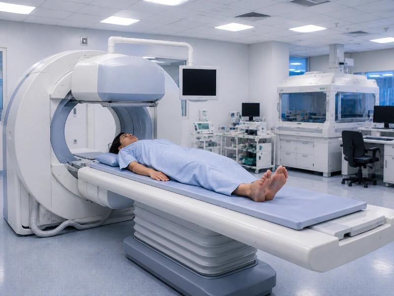
A Renal EC Scan evaluates individual kidney blood supply, active tubular secretion, and structural urinary drainage patterns. Clinically known as a Technetium-99m Ethylenedicysteine (Tc-99m EC) renogram, this procedure targets extraction mechanisms across your renal tissues. While other tests focus strictly on passive filtering, the EC scan checks how rapidly your tubules pull waste from your blood. Therefore, our imaging facility utilizes advanced automated gamma cameras to record fluid kinetics. This high-precision tracking measures effective renal plasma flow (ERPF) and split-renal differential percentages with exceptional clinical accuracy.
Furthermore, the EC radiotracer features an exceptionally high extraction rate across renal structures. Consequently, it delivers remarkably sharp visual outlines of your functional paths, even in patients with severely reduced kidney output. This precision makes it the absolute gold-standard choice for evaluating childhood urinary tracts or managing advanced chronic scarring. However, capturing these delicate drainage curves requires calibrated tracking arrays and expert interpretation. For this reason, our dedicated hubs use standardized nuclear software. As a result, nephrologists and pediatric surgeons can isolate mechanical drainage blocks and build accurate treatment pathways.
What is a Comprehensive Renal EC Scan?
A renal EC scan is a dynamic functional imaging profile that relies on the active tubular secretion properties of technetium-labeled ethylenedicysteine. Therefore, precise laboratory calibration allows our nuclear medicine physicians to chart arterial blood flow, calculate individual organ clearing percentages, and evaluate urinary drainage paths for a complete renal assessment.
- Perfusion Phase (Evaluating real-time blood movement to the left and right kidneys)
- Tubular Extraction Phase (Calculating precise split-renal differential percentages and plasma flow)
- Excretory and Clearance Phase (Mapping out how quickly the tracer exits into the bladder)
- Diuretic Challenge (Lasix) Phase (Using specialized prompts to differentiate physical blocks from simple slow pooling)
Renal EC Scan Diagnostic Pricing Guide
To ensure complete financial transparency for our patients, we provide clear, upfront cost structures across our advanced nuclear medicine imaging hubs. Below is the current price breakdown for renal EC imaging variations and associated biochemical panel matchings at our Navi Mumbai and Mumbai centers:
| Diagnostic Scan / Panel Variation | Estimated Cost (INR) |
|---|---|
| Standard Renal EC Scan (with Split Function) | ₹6,000 – ₹7,500 |
| Diuretic Renal EC Scan (with Lasix Injection) | ₹7,000 – ₹8,500 |
| Comprehensive Tubular Evaluation Panel | ₹8,200 – ₹9,800 |
Note: Final pricing variations can occur based on specialized pharmaceutical requirements or specific clinical instructions provided by your referring consultant.
Why is a Renal EC Scan Performed?
Physicians commonly recommend a dynamic EC kidney scan when evaluating children, checking structural performance in poorly functioning or damaged kidneys, or when investigating suspected urinary tract blocks. Moreover, the analytical findings directly assist urologists, pediatric surgeons, and transplant teams in choosing the safest treatment paths. Our experienced clinical team ensures you receive highly accurate imaging and reports.
- Calculating accurate split functional percentages in damaged or poorly functioning kidneys
- Investigating suspected Hydronephrosis (swelling of the kidney from built-up urine)
- Differentiating a true mechanical block (like a stone or stricture) from a dilated but clear drainage tube
- Evaluating kidney health in infants under 3 months of age, where tubular testing provides superior clarity
- Routine performance checking and clearance surveillance of a transplanted kidney
Conditions Diagnosed and Monitored via EC Scintigraphy
An accurate dynamic EC scan helps clinicians identify, track, and manage a wide variety of structural, functional, and vascular kidney disorders. You can find detailed clinical guidelines from the National Kidney Foundation standards.
- Ureteropelvic Junction (UPJ) Obstruction (Blocks where the kidney meets the drainage tube)
- Renal Tubular Dysfunction and chronic interstitial tissue changes
- Hydronephrosis and chronic urinary pooling tracking
- Renal Scarring (Evaluating permanent tissue damage from childhood UTIs)
If your dynamic kidney scan reveals filtering drops, your physician can cross-reference your results with other key diagnostic checks. For example, you can review your blood waste levels and mineral markers with our Kidney Function Test services or our comprehensive blood test services. Additionally, you can check blood cell balances through our Complete Blood Count (CBC) test services or our targeted HbA1c test services. We also offer standard Liver Function Test services, a comprehensive Lipid Profile test, a routine Urine Test, and neuroendocrine master mapping with our specialized MRI Pituitary Brain services. Alternatively, if you need standard samples collected comfortably at home while resting, you can book a certified mobile professional through our home blood collection services.
Preparation for a Renal EC Scan
- Fasting is typically not required for a standalone renal scan; you can eat a light meal normally before your appointment.
- Drink plenty of fluids—at least 3 to 4 glasses of plain water—in the 1 hour before your scan for clearance accuracy.
- Inform our laboratory staff about all daily medications, blood pressure prescriptions, or diuretics you take.
- Diuretics (water pills) should generally be paused on the day of the scan unless requested by your doctor.
- Let our imaging team know if you are pregnant, nursing, or have a history of severe allergies to nuclear medicine tracers.
What to Expect During the Imaging Process
- Our professional nuclear medicine team registers you and reviews your kidney care history in our imaging suite.
- A technician checks your hydration level and may ask you to drink another glass of plain water.
- You will lie comfortably flat on your back on a cushioned imaging table, with the camera positioned directly around your lower back.
- A small intravenous (IV) line is placed into a vein in your arm, and the safe, low-dose EC tracking agent is injected through it.
- The gamma camera immediately starts recording dynamic frames, and you must lie completely still for 25 to 30 minutes.
- If a diuretic check is needed, the Lasix injection is introduced through the IV halfway through the scan to record your drainage response.
The entire imaging sequence is highly efficient, typically requiring less than an hour of continuous scanning to complete safely.
Frequently Asked Questions
What is the main difference between a DTPA scan and an EC scan?
A DTPA scan enters the kidneys strictly through passive glomerular filtration, making it the preferred choice for measuring GFR. In contrast, an EC scan enters the kidneys through active tubular secretion. Because the kidneys extract the EC tracer much faster, it provides much sharper images and more reliable functional curves in damaged kidneys or young children.
Is the EC tracking injection painful, and are there any side effects?
The scan itself is completely painless. You will only feel a standard mild pinch when the IV line is placed in your arm. The EC tracer uses a very low, safe dose of diagnostic radiation that leaves your body naturally through your urine within 24 hours. It does not cause drowsiness or nausea, meaning you can drive home right after the test.
Why is a renal EC scan highly preferred for pediatric patients?
In infants under 3 months of age, the passive filtration system is not yet fully developed, which can make DTPA results difficult to read. Specifically, the active clearance paths mature much faster. Therefore, a renal EC scan delivers significantly clearer images and highly dependable diagnostic data for young children.
How long does it take to get my verified kidney scan reports?
Our nuclear medicine facility processes your dynamic flow charts and split tubular function curves efficiently. As a result, a senior specialist reviews the clearance metrics, and a verified interpretation report is usually ready for your doctor within 24 hours.
Why is a nuclear renal scan preferred over waiting out back or side pain?
Ultimately, a hidden urinary tract block or a silent drop in individual kidney function can cause long-term, irreversible tissue scarring if left unmanaged. High-precision EC scintigraphy is the only definitive way to measure your live tubular clearance, remove the medical guesswork, and protect your long-term health safely.
Why Our Diagnostics Laboratory is the Premier Choice for Renal Imaging
Our medical center remains the premier choice for monitoring your health because we combine high-resolution gamma camera technology with advanced split tubular function software. Furthermore, our clinical workflows eliminate tracking anomalies, our quality control protocols align with top pathology standards, and we ensure rapid delivery of your reports so you and your doctor stay fully informed. This steady focus on diagnostic accuracy makes our imaging facility a trusted partner in protecting your long-term renal, vascular, and metabolic wellness.