Dopa Scan: Fast & Precise F-18 DOPA PET CT Imaging
First of all, a Dopa Scan (F-18 Fluorodopa PET CT) represents the premier diagnostic imaging tool for assessing presynaptic dopaminergic pathways in movement disorders and evaluating neuroendocrine tumors. Essentially, this advanced molecular scan measures L-dopa decarboxylase activity and dopamine transporter density in the brain’s basal ganglia. As a result, neurologists and oncologists achieve unmatched diagnostic precision.
Book your Fluorodopa F 18 PET scan at Henotic Diagnostics for cutting-edge nuclear medicine clarity. Furthermore, our accredited diagnostic centre operates 24/7 to provide seamless appointment scheduling.
📋 Your Dopa Scan at a Glance
| 🔍 Detail | 📋 Info |
|---|---|
| 🧬 Radiotracer | Fluorine-18 Fluorodopa (F-18 DOPA) Radiotracer |
| ⏱️ Procedure Time | 2 to 3 Hours (Includes tracer uptake period) |
| 💰 Price Range | ₹20,000 – ₹26,000 (All-inclusive diagnostic fee) |
| 📄 Report TAT | 12–24 Hours (WhatsApp + Digital Portal + Hardcopy) |
| 🕐 Availability | 24/7 Operations (Prior slot reservation required) |
| 👩⚕️ Specialist | Dr. Pratibha Patil — Chief Consultant Radiologist |
| 🍽️ Fasting | 4 to 6 Hours Fasting required before appointment |
| 💳 Insurance | Cashless & Reimbursement Support available |
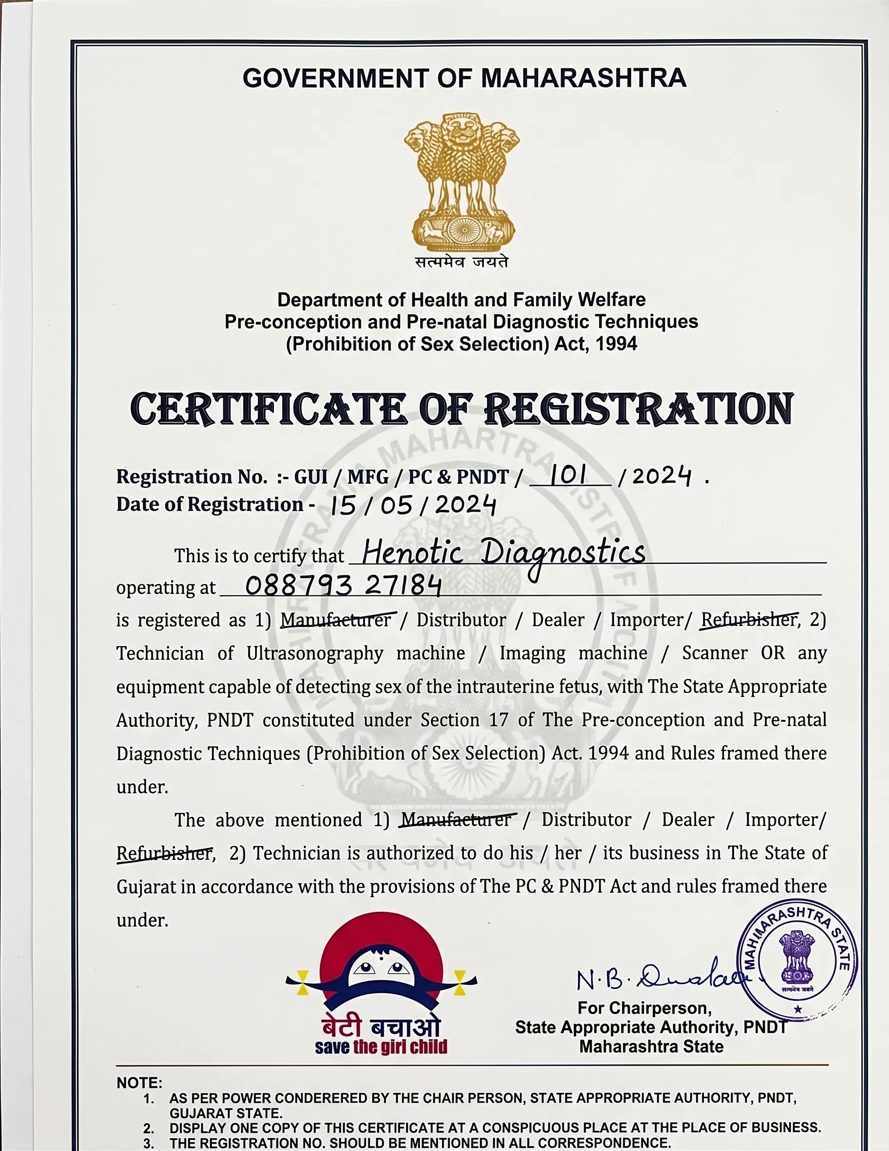
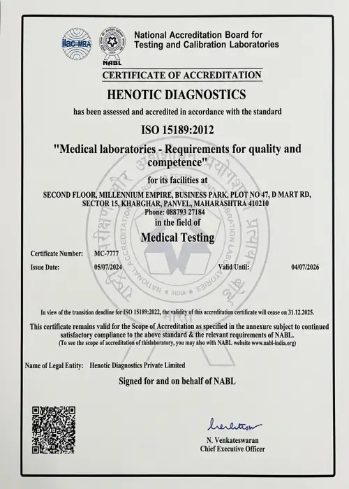
| 🏅 Accreditation | NABL Accredited · PCPNDT Certified · AERB Compliant |
Dopa Scan Quality Standards & Certifications
Overview of Dopa Scan PET CT Molecular Imaging
To begin with, a Fluorodopa F 18 PET scan provides unmatched molecular visualization of amino acid transport and dopaminergic metabolism in the human body. At the same time, specialists at Henotic Diagnostics integrate high-resolution PET CT technology with Fluorine-18 labeled L-DOPA radiotracers.
What Does the Scan Evaluate?
Specifically, the scan evaluates striatal presynaptic dopaminergic loss in Parkinson’s disease, differentiated neuroendocrine tumors, pheochromocytomas, and congenital hyperinsulinism. Consequently, neurologists and oncologists rely on these metabolic maps to differentiate idiopathic Parkinson’s from essential tremor. Furthermore, high-precision imaging identifies catecholamine-secreting tumors. As a result, physicians can plan optimal medical or surgical interventions. In fact, to review clinical guidelines, consult the RadiologyInfo PET resource.
 Furthermore, state-of-the-art Fluorodopa F 18 PET scan imaging delivers high sensitivity dopaminergic mapping
What is a Dopa Scan?
In general, a Fluorodopa F 18 PET scan is a specialized PET CT imaging procedure using Fluorine-18 L-dihydroxyphenylalanine (F-18 DOPA). Essentially, F-18 DOPA acts as an analogue of natural L-DOPA, accumulating in dopaminergic nerve terminals and neuroendocrine tumor cells. Specifically, when injected intravenously, it crosses the blood-brain barrier into the striatum (putamen and caudate nucleus). Therefore, 3D PET CT fusion scanners capture early loss of dopamine transporters before severe clinical motor symptoms emerge. Additionally, this diagnostic sensitivity far surpasses conventional structural MRI or SPECT imaging.
 Additionally, F-18 DOPA radiotracer uptake enables precise basal ganglia evaluation
What Does a Dopa Scan Check?
Overall, the scan provides full neuro-functional and whole-body evaluation based on your primary referral indication:
Key Anatomical & Neurological Structures Examined
 In addition, 3D PET CT fusion images pinpoint dopamine decarboxylase expression
When Is This Test Recommended?
For example, doctors recommend a Fluorodopa F 18 PET scan for clear clinical indications in neurology and endocrinology. In particular, neurologists order the scan to differentiate Parkinson’s disease from essential tremor, progressive supranuclear palsy, or vascular parkinsonism. Moreover, oncologists request this scan for staging medullary thyroid cancer and glomus tumors. In addition, nuclear medicine specialists use this scan to evaluate recurrent cerebral gliomas.
 Moreover, expert diagnostic evaluation guarantees comprehensive reporting and staging
Emergency Warning Symptoms for Neurological & Endocrine Health
Ideally, you should consult your specialist immediately if you experience rapid motor deterioration, unprovoked severe hypertensive crises, or acute neurological deficits. In addition, if severe tremors accompany postural instability, prompt medical assessment is vital. Indeed, early diagnostic scanning enables timely therapeutic adjustments.
Dopa Scan Cost Guide
| Nuclear Medicine Service | Approximate Cost (₹) |
|---|---|
| F-18 Dopa Scan (Brain / Dopaminergic) | First, F-18 Dopa Scan pricing is ₹20,000 – ₹24,000. |
| F-18 Dopa Whole Body PET Scan (NET / Pheo) | Second, whole body Dopa PET CT costs ₹22,000 – ₹26,000. |
| Whole Body FDG PET CT Scan | In addition, general FDG PET CT scan costs ₹12,000 – ₹16,000. |
| Nuclear Medicine Specialist Consultation | Finally, expert consultation fee is ₹1,000. |
Comparison: Dopa Scan vs. Conventional MRI & DAT SPECT
On one hand, structural MRI shows normal brain anatomy in early Parkinson’s disease, whereas an F-18 Dopa Scan detects early biochemical presynaptic deficits in the posterior putamen. Consequently, Fluorodopa F 18 PET scan provides superior resolution compared to conventional SPECT imaging.
| Feature | F-18 Dopa Scan PET CT | Brain MRI / DAT SPECT |
|---|---|---|
| Diagnostic Target | First, Dopa Scan measures dopamine decarboxylase activity. | However, MRI measures anatomical brain structure only. |
| Spatial Resolution | Second, 3D PET CT offers sub-4mm spatial resolution. | In contrast, DAT SPECT resolution is 8–10mm. |
| Early Parkinson Detection | In addition, Fluorodopa F 18 PET scan detects early asymmetrical putaminal loss. | Meanwhile, early Parkinson MRI scans appear completely normal. |
| Neuroendocrine Utility | Finally, Fluorodopa F 18 PET scan evaluates NETs, pheochromocytomas & glioblastomas. | Otherwise, conventional brain MRI cannot evaluate whole-body NETs. |
Frequently Asked Questions
Is a Dopa Scan painful? ▼
However, you experience only a minor intravenous needle stick during radiotracer administration, while the scanning process is entirely painless.
Do I need to fast before a Dopa Scan? ▼
Because metabolic radiotracer uptake must be optimized, fasting for 4 to 6 hours before the appointment is recommended.
How long does the appointment take? ▼
Generally, the complete procedure takes approximately 2 to 3 hours including radiotracer uptake time.
How long until I receive diagnostic results? ▼
Fortunately, detailed nuclear medicine diagnostic reports are provided within 12 to 24 hours.
What is the cost of a Dopa Scan? ▼
Overall, a high-precision F-18 Dopa Scan ranges from ₹20,000 to ₹26,000 all-inclusive.
Quick Appointment Booking
Schedule your Fluorodopa F 18 PET scan with accredited radiodiagnostic experts.
Call: 088793 27184 WhatsApp BookingScan Summary
Reporting Nuclear Medicine Specialist

Dr. Pratibha Patil
Chief Consultant Radiologist & Nuclear Medicine Specialist
MBBS, MD (Radiodiagnosis) · 15+ Years Clinical Experience
Furthermore, your Dopa Scan images are reviewed and interpreted by senior radiodiagnostic professionals. Consequently, comprehensive reports provide neurology and oncology teams with precise diagnostic data to optimize treatment planning.
Our Accreditations & Quality Standards
Certified excellence ensuring safety, radiation compliance, and accurate diagnostic results.




What Our Patients Say
Real feedback from patients who completed Dopa Scan PET CT imaging with us.
“Dr. Pratibha Patil’s F-18 DOPA scan report differentiated my father’s Parkinson’s symptoms clearly. Furthermore, the report was delivered within 24 hours.”
– Suresh B.
Verified Patient Review“The Dopa Scan identified my neuroendocrine lesion location with extreme clarity. In addition, the imaging team was exceptionally supportive throughout.”
– Radhika P.
Verified Patient Review“High quality PET CT scanner, clean environment, and transparent pricing. My neurologist was very satisfied with the image resolution.”
– Mohan K.
Verified Patient ReviewRelevant Diagnostic PET CT & Nuclear Medicine Services
Corporate Booking Office & Location
If you require a high precision Dopa Scan for Parkinson’s or neuroendocrine tumor evaluation, do not delay. Call to schedule your appointment.
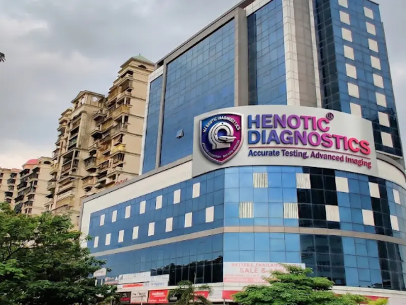
Henotic Diagnostics
Second floor, Millennium Empire, Business Park, Plot No 47, D Mart Rd, Sector 15, Kharghar, Navi Mumbai, Panvel, Maharashtra 410210
Phone: 088793 27184 | Hours: Open 24 Hours