MRI Enterography: High-Resolution 3T Small Bowel Imaging
First of all, an MRI Enterography scan provides a non-invasive, high-resolution diagnostic evaluation of the small intestine, including the duodenum, jejunum, ileum, and terminal ileocecal region. Essentially, this advanced 3T small bowel MR enterography (MRE) scan utilizes oral hyperosmolar contrast agents and IV gadolinium contrast to achieve maximal luminal distension and soft tissue contrast without radiation. As a result, gastroenterologists accurately diagnose Crohn’s disease activity, transmural bowel inflammation, fibrostenotic strictures, entero-enteric fistulae, mesenteric hypervascularity (comb sign), small bowel neoplasms, and obscure gastrointestinal bleeding.
Furthermore, you can book your MRI Enterography scan at Henotic Diagnostics for complete clinical precision and patient comfort. In addition, our accredited diagnostic facility operates 24/7 to provide prompt, seamless appointment booking.
Your MRI Enterography Scan Summary at a Glance
| First, Diagnostic Detail | Second, Clinical Information |
|---|---|
| First, Test Parameter | First, High-Resolution 3T MRI Scan of Small Intestine & Mesenteric Vessels |
| Second, Procedure Time | Second, 45 to 60 Mins Drinking + 30 Mins Scan Time (Includes oral PEG/Mannitol solution) |
| In addition, Price Range | In addition, ₹9,500 – ₹11,500 (All-inclusive 3T MRI Enterography protocol) |
| Furthermore, Report TAT | Furthermore, 2 to 4 Hours (Full DICOM digital images + expert radiologist report) |
| Moreover, Availability | Moreover, 24/7 Operations (Prior slot reservation recommended) |
| Also, Specialist | Also, Dr. Pratibha Patil — Chief Consultant Radiologist & Diagnostics |
| Additionally, Fasting | Additionally, 6 Hours Fasting Required prior to arriving at the diagnostic center |
| Besides, Insurance | Besides, Cashless & Reimbursement Support available |
| Finally, Accreditation | Finally, NABL Accredited · PCPNDT Certified · AERB Compliant |
MRI Enterography Quality Standards and Certifications
Overview of MRI Enterography Scanning for Small Bowel & Abdominal Health
To begin with, an MRI Enterography scan delivers vital diagnostic insights when evaluating Inflammatory Bowel Disease (IBD), Crohn’s disease, unexplained abdominal pain, chronic diarrhea, bowel strictures, and intestinal bleeding. At the same time, specialists at Henotic Diagnostics utilize multi-channel 3T magnetic resonance scanners with specialized abdominal phased-array coils to generate sub-millimeter cross-sectional images of distended small bowel loops, mesenteric blood vessels, lymph nodes, and perianal structures.
What Does an MRI Enterography Scan Evaluate?
Specifically, the scan evaluates small bowel wall thickening, mucosal hyperenhancement, submucosal edema (halo sign), mesenteric hypervascularity (comb sign), skip lesions in Crohn’s disease, fibrostenotic luminal narrowing, pre-stenotic dilation, entero-enteric or entero-cutaneous fistulae, intra-abdominal abscesses, gastrointestinal stromal tumors (GIST), carcinoid tumors, and small bowel lymphoma. Consequently, gastroenterologists, colorectal surgeons, and IBD specialists rely on high-resolution T1, T2, STIR, Diffusion-Weighted Imaging (DWI), dynamic contrast-enhanced (DCE), and cine-MRI sequences to distinguish active inflammatory strictures from chronic fibrotic scar tissue cleanly. Furthermore, multiplanar MR images evaluate the full length of the small intestine without endoscopic access limits. In fact, to review abdominal enterography radiology guidelines, consult the RadiologyInfo resource.
 Furthermore, private 3T MRI scanning suite ensures patient comfort and high diagnostic precision.
What is an MRI Enterography Scan?
In general, an MRI Enterography scan is a specialized diagnostic imaging procedure combining oral bowel distension solutions (such as PEG or Mannitol) with high-field 3T magnetic resonance imaging. Essentially, drinking 1 to 1.5 liters of neutral oral contrast agent over 45 to 60 minutes distends the small intestine loops. Then, IV contrast gadolinium and anti-peristaltic medication (such as Buscopan or Glucagon) are administered to freeze bowel movement and highlight inflammatory vascularity. Therefore, physicians observe whether intestinal symptoms are caused by active Crohn’s disease, transmural ulcers, fistulous tracts, or neoplastic strictures without subjecting young IBD patients to ionizing radiation.
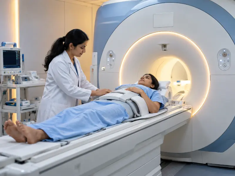 Additionally, wide-bore 3T MRI scanners deliver exceptional signal resolution and patient comfort.
Key Diagnostic Parameters & Findings in MRI Enterography Scanning
Overall, radiologists analyze four key structural and pathological parameters on multiplanar 3T MRI Enterography sequences:
MRI Enterography Key Clinical Indicators Examined
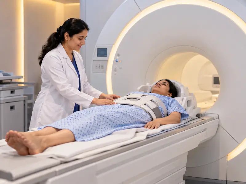 In addition, real-time multiplanar 3T MRI enterography scans accurately map Crohn’s inflammation, strictures, and fistulae.
When Is an MRI Enterography Scan Recommended?
For example, gastroenterologists, colorectal surgeons, and internists order an MRI Enterography scan when patients present with established or suspected Crohn’s disease, chronic right lower quadrant abdominal pain, unexplained weight loss, chronic diarrhea, elevated fecal calprotectin, suspected bowel obstruction, or gastrointestinal bleeding of obscure origin. In particular, doctors request testing to monitor response to biologic therapies (such as infliximab or adalimumab) and to differentiate active inflammatory strictures from fixed fibrotic scars before surgery. Moreover, it is the preferred imaging modality in young patients who require repeated bowel evaluations.
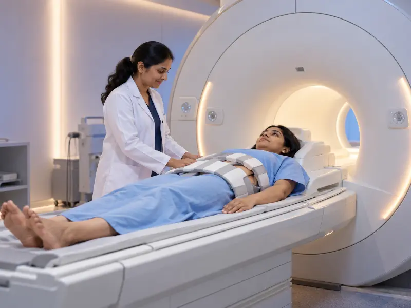 Moreover, high-resolution 3T MRI Enterography scanning is recommended for Crohn’s disease, IBD flares, and stricture evaluations.
Step-by-Step Patient Preparation and Prerequisites
First of all, proper preparation is essential for a successful MRI Enterography scan. Arrive 60 minutes before your appointment to consume the required oral contrast solution under radiologic supervision.
Fasting and Contrast Injection Conditions
In general, an MRI Enterography scan requires both oral contrast distension and intravenous gadolinium injection. Therefore, specific clinical protocols apply to ensure diagnostic precision:
Contrast Gadolinium Safety Protocol:
- First, maintain 6 hours of strict fasting prior to oral contrast and IV contrast administration.
- Second, provide recent Serum Creatinine / eGFR lab results to confirm normal kidney clearance.
- In addition, an IV cannula is placed safely into an arm vein for gadolinium injection and IV Buscopan anti-peristaltic administration.
- Furthermore, contrast gadolinium sharply highlights hyperenhancing inflamed bowel mucosa, active fistulae, and hypervascular mesenteric vessels.
What to Expect During the Procedure
Overall, the MRI Enterography scan is non-invasive, comfortable, and zero-radiation. After completing the oral contrast drinking phase, you lie comfortably on the padded scanner table prone or supine, with a multi-channel abdominal phased-array coil placed across your abdomen. As the table enters the wide-bore scanner, noise-reducing headphones buffer magnet tapping sounds. Short breath-holds (10-15 seconds) are requested during image acquisition. An IV injection of Buscopan is administered to temporarily pause bowel movement. The scanning process takes 25 to 35 minutes. Additionally, an emergency squeezable bulb is in your hand at all times for instant technologist contact.
Advanced Medical Technology: Machines and Models
At Henotic Diagnostics, we utilize state-of-the-art 3 Tesla (3T) wide-bore MRI systems engineered by Siemens and GE Healthcare. Specifically, our machines feature high-density body matrix coils and advanced pulse sequences (T1 VIBE, T2 HASTE, TrueFISP cine-MRI, STIR fat-suppression, and Diffusion-Weighted Imaging DWI). Therefore, our scanners resolve sub-millimeter mural thickening, subtle sinus tracts, and early mucosal enhancement that low-field scanners miss.
Post-Procedure Care & Aftercare Guidelines
Fortunately, recovery after an MRI Enterography scan is fast and straightforward. You may resume normal activities and return home following the exam.
Diagnostic Reporting and Hardcopy Availability
In addition, our digital workflow delivers fast, high-precision reporting. Following your scan, complete DICOM image sets are sent to our PACS server. Chief Radiologist Dr. Pratibha Patil evaluates axial, coronal, and sagittal cross-sections, delivering detailed diagnostic reports within 2 to 4 hours. Furthermore, printed laser hardcopy films and digital USB/cloud report links are available at reception.
Second Opinion & Subspecialty Consultations
Besides routine reporting, complex Crohn’s strictures, fistulizing disease, or surgical planning require expert subspecialty review. At Henotic Diagnostics, we provide dedicated second opinions and direct peer-to-peer case discussions with treating gastroenterologists and colorectal surgeons:
Subspecialty Abdominal Radiology Services:
- First, expert gastrointestinal radiology second opinion reviews on external MRE films or DICOM data.
- Second, direct doctor-to-doctor consultations between our radiologist and your attending gastroenterologist.
- In addition, precise differentiation between active inflammatory strictures vs fixed fibrotic scar tissue.
- Furthermore, instant encrypted digital image sharing for emergency clinical reviews.
Why Choose Us for Your Care?
Choosing Henotic Diagnostics for your MRI Enterography scan guarantees top-tier technology and compassionate care:
Transportation & Center Accessibility
Moreover, our diagnostic facility in Kharghar, Navi Mumbai, is designed for smooth accessibility:
Emergency Warning Symptoms for Bowel Health
Ideally, you should seek urgent medical evaluation if you experience acute severe abdominal pain, persistent vomiting with inability to pass gas or stool (complete bowel obstruction), high fever with severe abdominal tenderness (suspected bowel perforation or abscess), or heavy rectal bleeding. In addition, immediate diagnostic scanning is vital to guide emergency surgical intervention.
MRI Enterography Test Cost Guide
| First, Diagnostic Enterography Service | Second, Approximate Cost (₹) |
|---|---|
| First, Standard 3T MRI Enterography Scan (With Oral Contrast) | First, standard MRE scan cost is ₹9,500 – ₹11,500. |
| Second, Comprehensive Dynamic Contrast 3T MRI Enterography | Second, Contrast MRE with IV Gadolinium costs ₹14,500 – ₹16,500. |
| In addition, Combined MRI Enterography + MRI Pelvis Fistulogram | In addition, Combined MRE & Perianal Fistulogram costs ₹16,500 – ₹18,500. |
| Finally, Diagnostic Radiology Consultation | Finally, expert radiologist consultation fee is ₹1,000. |
Comparison: CT Enterography / Endoscopy vs. MRI Enterography
On one hand, Endoscopy and Colonoscopy evaluate the mucosal surface of the stomach, terminal ileum, and colon directly but cannot reach deep mid-small bowel loops or evaluate transmural wall thickness. CT Enterography (CTE) offers fast imaging but exposes young IBD patients to ionizing radiation. On the other hand, an MRI Enterography scan provides 100% zero-radiation transmural soft tissue imaging, easily distinguishing active inflammation from fibrotic strictures while assessing fistulae.
| First, Feature | Second, CT Enterography / Endoscopy | Third, MRI Enterography (3T MRE Scan) |
|---|---|---|
| First, Primary Strength | First, Endoscopy allows superficial biopsies; CTE is fast for trauma. | However, MRE excels at deep small bowel transmural wall & fistulae mapping. |
| Second, Radiation Safety in Young IBD Patients | Second, CTE delivers cumulative radiation over repeated follow-up scans. | In contrast, MRE uses magnetic fields with 100% zero radiation. |
| In addition, Active Inflammatory vs Fibrotic Differentiation | In addition, CT has limited soft tissue resolution for fibrotic stricture grading. | Meanwhile, MRE T2/DWI sequences cleanly distinguish active edema from fibrosis. |
| Finally, Deep Small Intestine Access | Finally, standard colonoscopes cannot reach mid-jejunum or upper ileum. | Otherwise, gold standard for 100% complete small intestine visualization. |
Frequently Asked Questions
First, Is an MRI Enterography scan painful? ▼
However, the scan is completely non-invasive and painless because it uses magnetic fields rather than invasive procedures.
Second, Why do I need to drink oral contrast before MRI Enterography? ▼
Because collapsed small bowel loops obscure mucosal details, drinking 1 to 1.5 liters of oral PEG solution distends the intestinal walls for accurate inflammation grading.
In addition, How long does the MRI Enterography scan take? ▼
Generally, oral contrast drinking takes 45 to 60 minutes, while the actual scanner acquisition takes 25 to 35 minutes.
Furthermore, When will I receive my MRI Enterography report? ▼
Fortunately, full DICOM digital scans and expert radiologist diagnostic reports are delivered within 2 to 4 hours.
Finally, What is the cost of an MRI Enterography scan? ▼
Overall, a standard 3T MRI Enterography scan ranges from ₹9,500 to ₹11,500 all-inclusive depending on contrast injection and dedicated protocol requirements.
Quick Appointment Booking
Schedule your MRI Enterography scan with accredited radiodiagnostic experts.
First, Call: 088793 27184 Second, WhatsApp BookingQuick Test Summary Overview
Reporting Diagnostic Specialist

Dr. Pratibha Patil
First, Chief Consultant Radiologist & Diagnostic Specialist
Second, MBBS, MD (Radiodiagnosis) · 15+ Years Clinical Experience
Furthermore, your MRI Enterography scans and multiplanar reconstructions are analyzed by senior gastrointestinal radiologists specializing in Inflammatory Bowel Disease and small bowel imaging. Consequently, comprehensive reports provide gastroenterologists and colorectal surgeons with precise anatomical data to evaluate Crohn’s disease activity, stricture characterization, and fistulating tracts.
Our Accreditations and Quality Standards
In addition, certified excellence ensures safety, hygiene compliance, and accurate diagnostic results.


What Our Patients Say
Furthermore, read real feedback from patients who completed MRI Enterography scanning with us.
“First of all, I suffered severe Crohn’s disease symptoms and needed an MRI Enterography scan. Dr. Pratibha Patil’s team guided me through the oral PEG drinking protocol smoothly, and the report clearly highlighted active ileal inflammation.”
– Rahul N.
First, Verified Patient Review“In addition, the staff made sure I was comfortable during the oral contrast drinking phase and scan acquisition. Highly recommended for IBD patients!”
– Sneha K.
Second, Verified Patient Review“Furthermore, rapid diagnostic reporting within 3 hours. Printed laser films and digital DICOM links were provided right away for my gastroenterologist.”
– Vikas R.
Finally, Verified Patient ReviewRelevant Diagnostic Radiology and Gastrointestinal Services
Corporate Booking Office and Location
If you require a high precision MRI Enterography scan for Crohn’s disease, IBD evaluation, small bowel strictures, or intestinal fistulae, do not delay. In addition, call us today to schedule your appointment.
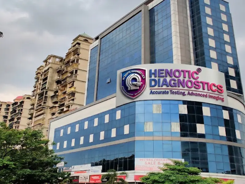
Henotic Diagnostics
First, Second floor, Millennium Empire, Business Park, Plot No 47, D Mart Rd, Sector 15, Kharghar, Navi Mumbai, Panvel, Maharashtra 410210
Phone: 088793 27184 | Hours: Open 24 Hours