MRI Orbit: High-Resolution 3T Optic Nerve & Ocular Imaging
First of all, an MRI Orbit scan provides a non-invasive, high-resolution diagnostic evaluation of retrobulbar soft tissue structures, including the optic nerve (CN II), optic sheath, extraocular muscles, lacrimal glands, intraconal and extraconal fat spaces, ocular globes, and cavernous sinuses. Essentially, this advanced 3T orbital MRI scan offers superior tissue contrast compared to CT imaging. As a result, radiologists accurately differentiate optic neuritis, thyroid eye disease (Graves’ ophthalmopathy), orbital pseudotumors, optic nerve sheath meningiomas, capillary hemangiomas, and orbital cellulitis.
Furthermore, you can book your MRI Orbit scan at Henotic Diagnostics for complete clinical precision and patient comfort. In addition, our accredited diagnostic facility operates 24/7 to provide prompt, seamless appointment booking.
Your MRI Orbit Scan Summary at a Glance
| First, Diagnostic Detail | Second, Clinical Information |
|---|---|
| First, Test Parameter | First, High-Resolution 3T MRI Scan of Orbits, Optic Nerves & Retrobulbar Spaces |
| Second, Procedure Time | Second, 25 to 45 Minutes (Includes axial, coronal, and sagittal T1/T2/STIR/DWI thin-slice sequences) |
| In addition, Price Range | In addition, ₹6,500 – ₹8,500 (All-inclusive 3T MRI Orbit scanning fee) |
| Furthermore, Report TAT | Furthermore, 2 to 4 Hours (Full DICOM digital images + expert radiologist report) |
| Moreover, Availability | Moreover, 24/7 Operations (Prior slot reservation recommended) |
| Also, Specialist | Also, Dr. Pratibha Patil — Chief Consultant Radiologist & Diagnostics |
| Additionally, Fasting | Additionally, 4 Hours Fasting for Contrast MRI (No fasting for non-contrast orbit MRI) |
| Besides, Insurance | Besides, Cashless & Reimbursement Support available |
| Finally, Accreditation | Finally, NABL Accredited · PCPNDT Certified · AERB Compliant |
MRI Orbit Quality Standards and Certifications
Overview of MRI Orbit Scanning for Ocular & Visual Health
To begin with, an MRI Orbit scan delivers vital diagnostic insights when evaluating sudden vision loss, painful eye movement, proptosis (eyeball protrusion), double vision (diplopia), and suspected optic nerve inflammation. At the same time, specialists at Henotic Diagnostics utilize multi-channel 3T magnetic resonance scanners with fat-saturated pulse sequences to generate sub-millimeter multiplanar cross-sectional images of the intraconal fat, optic sheath, extraocular muscles, and optic chiasm.
What Does an MRI Orbit Scan Evaluate?
Specifically, the scan evaluates optic neuritis (demyelination associated with Multiple Sclerosis or NMO), thyroid eye disease (extraocular muscle belly enlargement sparing tendons), orbital pseudotumor (idiopathic orbital inflammation), lacrimal gland tumors (pleomorphic adenomas, carcinomas), optic nerve sheath meningiomas, optic gliomas, cavernous hemangiomas, orbital cellulitis, and superior ophthalmic vein thrombosis. Consequently, ophthalmologists, neuro-ophthalmologists, neurologists, and ENT specialists rely on high-resolution T1, T2, STIR, Diffusion-Weighted Imaging (DWI), and contrast-enhanced images to characterize orbital pathology cleanly. Furthermore, multiplanar MR images distinguish inflammatory muscle edema from retrobulbar neoplasia. In fact, to review orbital radiology guidelines, consult the RadiologyInfo resource.
 Furthermore, private 3T MRI scanning suite ensures patient comfort and high diagnostic precision.
What is an MRI Orbit Scan?
In general, an MRI Orbit scan is a non-invasive diagnostic procedure that captures multiplanar magnetic resonance cross-sections of the entire orbital cavity, retrobulbar retro-ocular space, and optic nerve pathways. Essentially, radiofrequency pulses align hydrogen protons in optic nerve fibers, extraocular muscles, lacrimal tissue, and orbital fat, producing exceptionally detailed images without ionizing radiation. Therefore, physicians observe whether visual deficits are caused by optic nerve sheath enhancement, extraocular muscle compression, or retrobulbar masses. Additionally, this scan provides superior soft tissue resolution compared to ultrasound or CT.
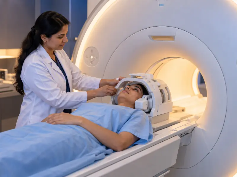 Additionally, wide-bore 3T MRI scanners deliver exceptional signal resolution and patient comfort.
Key Diagnostic Parameters & Findings in MRI Orbit Scanning
Overall, radiologists analyze four key structural and pathological parameters on multiplanar 3T MRI Orbit sequences:
MRI Orbit Key Clinical Indicators Examined
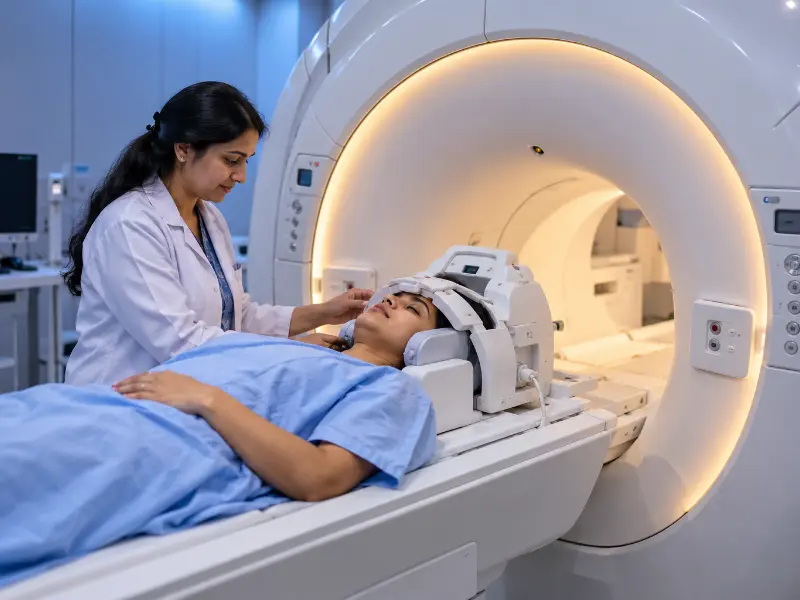 In addition, real-time multiplanar 3T MRI scans accurately map optic nerves, retrobulbar fat, and extraocular muscles.
When Is an MRI Orbit Scan Recommended?
For example, ophthalmologists, neurologists, and neuro-ophthalmologists order an MRI Orbit scan when patients present with painful eye movements, sudden onset vision blurriness or color loss, bulging eyes (proptosis), double vision (diplopia), or unexplained orbital swelling. In particular, doctors request testing prior to orbital decompression surgery, tumor biopsy, or when CT scans show ambiguous soft tissue involvement near the optic canal. Moreover, it is essential for diagnosing demyelinating conditions such as Multiple Sclerosis.
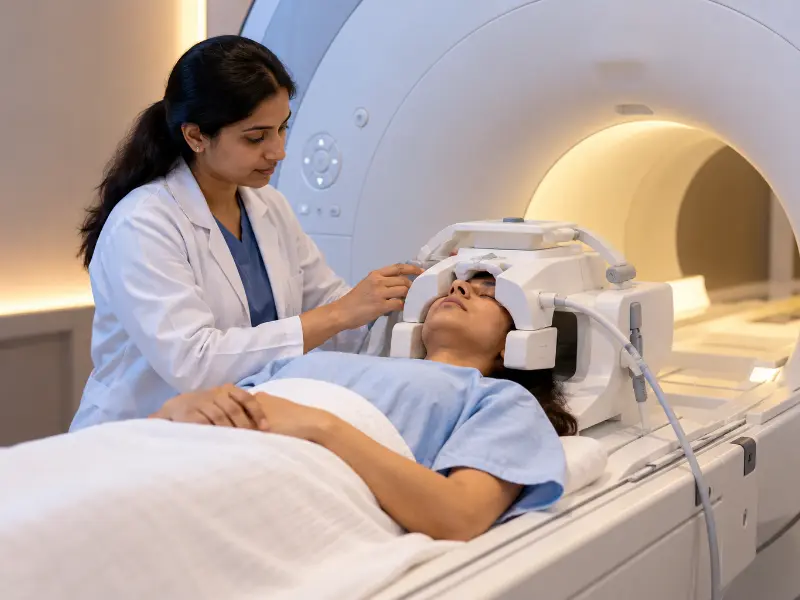 Moreover, high-resolution 3T MRI Orbit scanning is recommended for complex optic nerve, extraocular muscle, and retrobulbar evaluations.
Step-by-Step Patient Preparation and Prerequisites
First of all, proper preparation ensures a smooth MRI Orbit examination. Prior to entering the scanner room, patients complete a comprehensive safety screening checklist to identify any metallic implants, pacemakers, neurostimulators, or metallic foreign bodies.
Fasting and Contrast Injection Conditions
In general, non-contrast MRI Orbit scans require no fasting whatsoever. However, because contrast-enhanced MRI (CE-MRI Orbit) is frequently ordered to evaluate optic neuritis, orbital tumors, or inflammatory pseudotumors, specific clinical conditions apply:
Contrast Gadolinium Safety Protocol:
- First, maintain 4 hours of strict fasting (water permitted) prior to contrast administration.
- Second, provide recent Serum Creatinine / eGFR lab results to confirm normal kidney clearance.
- In addition, a small IV cannula is placed safely into an arm vein for gadolinium contrast injection during scanning.
- Furthermore, contrast gadolinium sharply delineates optic nerve sheath enhancement, vascular lesions, and orbital abscess capsule walls.
What to Expect During the Procedure
Overall, the MRI Orbit scan is painless, comfortable, and zero-radiation. You will lie on your back on the padded scanner table, with a specialized lightweight head-and-orbital coil positioned gently over your head. As the table moves into the wide-bore scanner, noise-reducing headphones are provided to buffer magnet tapping sounds. The scan takes 25 to 45 minutes, during which you simply lie comfortably, keep your eyes closed gently, and refrain from moving your eyes rapidly during specific short imaging sequences. Additionally, an emergency squeezable bulb is in your hand at all times for instant technologist contact.
Advanced Medical Technology: Machines and Models
At Henotic Diagnostics, we utilize state-of-the-art 3 Tesla (3T) wide-bore MRI systems engineered by Siemens and GE Healthcare. Specifically, our machines feature high-density orbital array coils and advanced fat-saturated pulse sequences (T1, T2, STIR fat-suppression, and Diffusion-Weighted Imaging DWI). Therefore, our scanners resolve sub-millimeter retrobulbar optic nerve lesions and detect early optic sheath enhancement that low-field scanners miss.
Post-Procedure Care & Aftercare Guidelines
Fortunately, recovery after an MRI Orbit scan is immediate. Once the scan is complete, you may resume normal activities, return to work, and drive home without any downtime.
Diagnostic Reporting and Hardcopy Availability
In addition, our digital workflow delivers fast, high-precision reporting. Following your scan, complete DICOM image sets are sent to our PACS server. Chief Radiologist Dr. Pratibha Patil evaluates axial, coronal, and sagittal cross-sections, delivering detailed diagnostic reports within 2 to 4 hours. Furthermore, printed laser hardcopy films and digital USB/cloud report links are available at reception.
Second Opinion & Subspecialty Consultations
Besides routine reporting, complex optic nerve sheath meningiomas, thyroid ophthalmopathy decompression planning, or retrobulbar masses require expert subspecialty review. At Henotic Diagnostics, we provide dedicated second opinions and direct peer-to-peer case discussions with treating neuro-ophthalmologists, neurologists, and orbital surgeons:
Subspecialty Neuro-Radiology & Orbital Services:
- First, expert neuroradiology second opinion reviews on external orbital MRI films or DICOM data.
- Second, direct doctor-to-doctor consultations between our radiologist and your attending neuro-ophthalmologist.
- In addition, multiplanar mapping of extraocular muscle belly enlargement prior to orbital decompression.
- Furthermore, instant encrypted digital image sharing for emergency clinical reviews.
Why Choose Us for Your Care?
Choosing Henotic Diagnostics for your MRI Orbit scan guarantees top-tier technology and compassionate care:
Transportation & Center Accessibility
Moreover, our diagnostic facility in Kharghar, Navi Mumbai, is designed for smooth accessibility:
Emergency Warning Symptoms for Orbital & Visual Health
Ideally, you should seek urgent medical evaluation if you experience sudden rapid vision loss (suspected acute optic neuritis), severe painful proptosis with high fever (suspected orbital cellulitis or abscess), acute painful ophthalmoplegia (inability to move the eye), or suspected cavernous sinus thrombosis. In addition, immediate 3T MRI Orbit scanning is vital to preserve optic nerve function and prevent intracranial complications.
MRI Orbit Test Cost Guide
| First, Diagnostic Orbital Service | Second, Approximate Cost (₹) |
|---|---|
| First, Standard Non-Contrast 3T MRI Orbit Scan | First, standard MRI Orbit scan cost is ₹6,500 – ₹8,500. |
| Second, Contrast-Enhanced 3T MRI Orbit Scan (CE-MRI) | Second, Contrast MRI Orbit costs ₹10,500 – ₹12,500. |
| In addition, Combined MRI Brain & Orbit 3T Scan | In addition, Brain & Orbit MRI costs ₹12,000 – ₹14,500. |
| Finally, Diagnostic Radiology Consultation | Finally, expert radiologist consultation fee is ₹1,000. |
Comparison: CT Scan Orbit vs. MRI Orbit
On one hand, CT Scan Orbit excels at demonstrating acute orbital bony fractures and metallic foreign bodies rapidly. On the other hand, an MRI Orbit scan provides superior soft tissue resolution, easily distinguishing optic nerve demyelination, extraocular muscle edema, intraconal tumor margins, and cavernous sinus invasion without radiation.
| First, Feature | Second, CT Scan Orbit | Third, MRI Orbit (3T Soft Tissue MRI) |
|---|---|---|
| First, Primary Strength | First, CT excels at acute bony fractures & radiopaque foreign objects. | However, MRI excels at optic nerve, retrobulbar fat & muscle detail. |
| Second, Optic Neuritis Demyelination | Second, CT cannot visualize intra-sheath optic nerve demyelination. | In contrast, MRI fat-sat STIR clearly shows optic nerve hyperintensity. |
| In addition, Radiation Exposure | In addition, uses low-dose ionizing X-ray radiation. | Meanwhile, uses magnetic fields with 100% zero radiation. |
| Finally, Cavernous Sinus & Apex Mapping | Finally, moderate soft tissue contrast near bony canals. | Otherwise, gold standard for cavernous sinus & intracranial extension. |
Frequently Asked Questions
First, Is an MRI Orbit scan painful? ▼
However, the scan is completely non-invasive and painless because it uses magnetic fields rather than invasive procedures.
Second, Do I need fasting before an MRI Orbit scan? ▼
Because non-contrast MRI requires no fasting, you may eat normally. However, 4 hours fasting is required if gadolinium contrast is administered.
In addition, How long does the MRI Orbit scan take? ▼
Generally, the complete scan takes 25 to 45 minutes depending on whether contrast-enhanced pulse sequences or combined brain protocols are required.
Furthermore, When will I receive my MRI Orbit report? ▼
Fortunately, full DICOM digital scans and expert radiologist diagnostic reports are delivered within 2 to 4 hours.
Finally, What is the cost of an MRI Orbit scan? ▼
Overall, a standard 3T MRI Orbit scan ranges from ₹6,500 to ₹8,500 all-inclusive depending on contrast injection and combined brain protocol requirements.
Quick Appointment Booking
Schedule your MRI Orbit scan with accredited radiodiagnostic experts.
First, Call: 088793 27184 Second, WhatsApp BookingQuick Test Summary Overview
Reporting Diagnostic Specialist

Dr. Pratibha Patil
First, Chief Consultant Radiologist & Diagnostic Specialist
Second, MBBS, MD (Radiodiagnosis) · 15+ Years Clinical Experience
Furthermore, your MRI Orbit scans and multiplanar reconstructions are analyzed by senior neuroradiologists specializing in ocular and neuro-ophthalmic imaging. Consequently, comprehensive reports provide ophthalmologists and neurologists with precise anatomical data to evaluate optic neuritis, thyroid eye disease, and retrobulbar masses.
Our Accreditations and Quality Standards
In addition, certified excellence ensures safety, hygiene compliance, and accurate diagnostic results.


What Our Patients Say
Furthermore, read real feedback from patients who completed MRI Orbit scanning with us.
“First of all, Dr. Pratibha Patil’s team provided a very clear 3T MRI Orbit report detecting early optic neuritis. Helped my neuro-ophthalmologist start treatment quickly!”
– Ananya V.
First, Verified Patient Review“In addition, the wide-bore MRI machine was very comfortable. The head coil was lightweight and staff made the whole procedure very easy.”
– Deepak R.
Second, Verified Patient Review“Furthermore, prompt report delivery within 2 hours for my orbital scan. Printed laser films and digital DICOM links were provided right away. Highly recommended facility.”
– Smita N.
Finally, Verified Patient ReviewRelevant Diagnostic Radiology and Ocular Services
Corporate Booking Office and Location
If you require a high precision MRI Orbit scan for optic nerve neuritis, thyroid eye disease, proptosis, or retrobulbar tumors, do not delay. In addition, call us today to schedule your appointment.
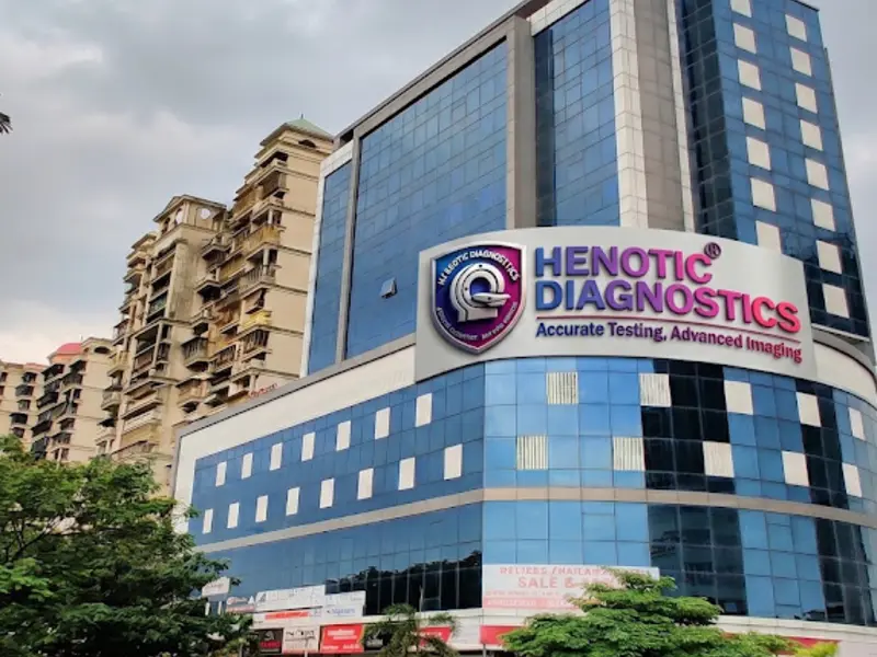
Henotic Diagnostics
First, Second floor, Millennium Empire, Business Park, Plot No 47, D Mart Rd, Sector 15, Kharghar, Navi Mumbai, Panvel, Maharashtra 410210
Phone: 088793 27184 | Hours: Open 24 Hours