Advanced Bilateral Sono Mammography (Breast Ultrasound)
Accurate, radiation-free, high-resolution breast ultrasound interpreted by certified senior consultant radiologists. Specifically, our women's imaging department provides gentle clinical examinations, same-day appointments, and fast WhatsApp report delivery within 2 hours.
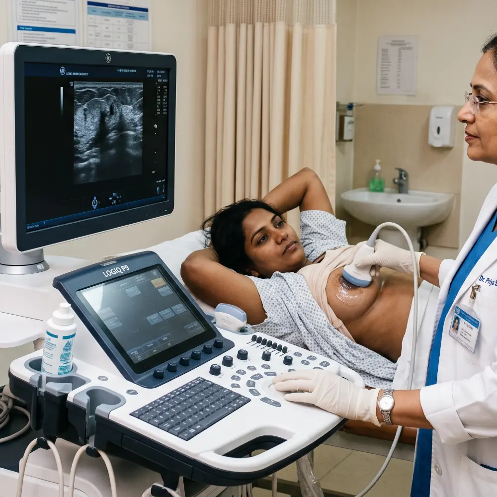
Sono Mammography at a Glance
Furthermore, review essential clinical details regarding the examination, scan timing, preparation requirements, and digital reporting protocols.
| Service Information | Details |
|---|---|
| Examination | Sono Mammography / Breast Ultrasound |
| Imaging method | Ultrasound (high-frequency sound waves) |
| Ionizing radiation | No radiation involved (completely safe) |
| Fasting required | Not required (eat and drink normally) |
| Preparation needed | Minimal (wear comfortable two-piece clothing) |
| Typical duration | Approximately 15–30 minutes |
| Patient experience | Generally well tolerated and painless |
| Interpretation | Senior Consultant Radiologist reporting |
| Digital reports | Yes (WhatsApp, Email & Patient Portal) |
| Hardcopy report | Available at center on request |
| Center availability | Open 24 hours for emergencies & scheduled scans |
| Appointment booking | Prior booking recommended for zero waiting |
What Is Sono Mammography?
Sono Mammography, also widely known as breast ultrasound, is an advanced non-invasive imaging examination specifically utilized to evaluate breast tissue architecture, palpable lumps, cysts, and suspicious masses.
During the examination, high-frequency acoustic sound waves generate real-time cross-sectional images of the breast parenchyma. Furthermore, our senior radiologists assess these live images to characterize tissue consistency and rule out underlying pathologies.
Specifically, Sono Mammography can effectively evaluate:
- Palpable breast lumps and localized breast pain
- Fluid-filled breast cysts and solid architectural masses
- Focal breast abnormalities detected during clinical exams
- Previously identified breast lesions requiring interval follow-up
- Dense breast tissue where X-ray mammograms may remain inconclusive
Consequently, unlike standard X-ray mammography, breast ultrasound utilizes zero ionizing radiation, making it completely safe for repeated evaluations.
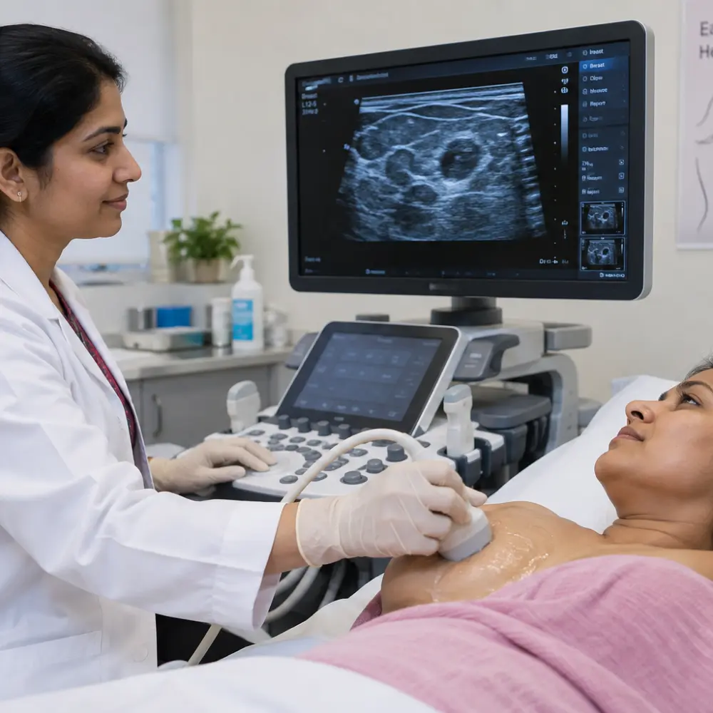
Why Is Sono Mammography Performed?
Clinicians recommend Sono Mammography whenever targeted anatomical information is required regarding a palpable finding, focal breast pain, or screening abnormality.
Evaluation of a New Lump
First, ultrasound provides instant differentiation between benign fluid-filled cysts and solid masses whenever a new breast lump is detected.
Dense Breast Tissue
In addition, dense glandular tissue can obscure small lesions on standard mammography, whereas ultrasound penetrates dense parenchyma effortlessly.
Focal Pain Assessment
Furthermore, patients experiencing localized tenderness receive real-time targeted sonographic scanning to inspect underlying ducts and lobules.
Evaluation During Pregnancy
Because ultrasound emits zero radiation, it remains the safest imaging modality for pregnant or lactating women needing breast evaluation.
Mammography Follow-up
Moreover, when a screening mammogram identifies an asymmetrical density, ultrasound provides complementary multi-angle morphological clarity.
USG-Guided Procedures
Finally, precision real-time sonography facilitates targeted needle guidance for fine needle aspiration cytology (FNAC) and core biopsies.
Sono Mammography vs Mammography: Understanding the Differences
While both examinations assess breast health, they rely on entirely different technologies and serve complementary diagnostic functions.
| Evaluation Feature | Sono Mammography (Breast Ultrasound) | X-Ray Mammography |
|---|---|---|
| Technology | High-frequency acoustic sound waves | Low-dose X-ray radiation |
| Ionizing Radiation | Zero radiation (100% safe) | Minimal low-dose radiation |
| Primary Strength | Differentiating fluid cysts from solid lumps | Detecting microcalcifications & structural distortion |
| Dense Breast Tissue | Highly effective in dense tissue | Sensitivity reduced by dense tissue overlay |
| Suitability in Young Women | First-choice modality for women under 40 | Generally recommended for women aged 40 and above |
| Compression Required | Gentle probe touch; no painful compression | Firm paddle compression required for image clarity |
Clinical Note: Importantly, Sono Mammography does not automatically replace mammography. In fact, clinicians frequently order both examinations concurrently to achieve maximum diagnostic sensitivity for comprehensive breast screening.
What Can Sono Mammography Evaluate?
Specifically, high-resolution sonography reveals intricate tissue morphology, enabling radiologists to classify findings accurately according to international BI-RADS criteria.
Breast Lumps & Solid Masses
Usually, a palpable lump can arise from various etiologies, the majority of which are benign fibroadenomas or simple cysts. Therefore, high-resolution ultrasound allows our radiologists to assess precise dimensions, internal vascularity via color Doppler, lesion borders, and acoustic transmission.
Breast Cysts & Fluid Collections
In addition, ultrasound excels at distinguishing simple benign cysts with thin walls from complex cysts requiring clinical surveillance. Consequently, patients receive immediate diagnostic clarity without undergoing invasive surgical biopsies unnecessarily.
| Ultrasound Assessment Metric | Clinical Diagnostic Significance |
|---|---|
| Cystic vs Solid Appearance | Instantly distinguishes fluid-filled benign cysts from cellular solid tumors. |
| Lesion Margins & Borders | Smooth circumscribed margins indicate benign etiology; irregular borders warrant closer evaluation. |
| Morphological Shape | Oval or wider-than-tall orientation typically correlates with benign fibroadenomas. |
| Internal Echo Texture | Anechoic, hypoechoic, or heterogeneous patterns provide critical histological clues. |
| Color Doppler Vascularity | Assesses blood flow patterns within the lesion to verify vascular activity. |
| Anatomical Quadrant Localization | Accurately documents clock-position coordinates and distance from the nipple. |
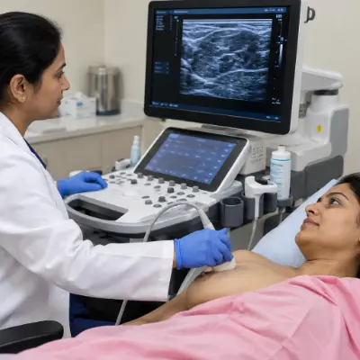
How Is Sono Mammography Performed?
Specifically, our breast imaging workflow is structured to ensure maximum comfort, complete dignity, and rigorous diagnostic accuracy.
Patient Registration
Initially, appointment confirmation, identity verification, and medical record intake are finalized at our desk.
Clinical History Review
Next, the radiology doctor discusses your symptoms, family history, and previous mammogram reports.
Private Positioning
Subsequently, you change privately into a sterile clinical gown and recline comfortably on the examination bed.
Gel Application
Then, warm, hypoallergenic ultrasound gel is applied to ensure seamless acoustic transmission.
Real-Time Scanning
Meanwhile, the radiologist glides the high-frequency probe across both breasts and axillary lymph nodes.
Image Acquisition
Following scanning, high-resolution static and Doppler video frames are captured for permanent records.
BI-RADS Reporting
Afterward, our certified consultant radiologist analyzes all images and issues a standardized report.
Rapid Delivery
Finally, your verified report is dispatched promptly on WhatsApp and email, with hardcopies available on request.
Our Senior Radiology Team
In fact, every Sono Mammography scan is interpreted and signed by senior consultant radiologists with decades of dedicated women's imaging expertise.

DR PRATIBHA PATIL
MBBS, MD (Radio-Diagnosis)
Specialist in Women's Imaging, Advanced Breast Ultrasound, BI-RADS Categorization, and Pelvic Sonography. Specifically, Dr. Patil has performed and supervised over 25,000 diagnostic imaging evaluations with exceptional clinical precision.

DR AMULYA PATIL
MBBS, DNB (Radio-Diagnosis)
Expert in High-Resolution Breast Ultrasound, Sono Mammography, Color Doppler hemodynamics, and USG-Guided FNAC interventions. Moreover, she is widely recognized for compassionate patient counseling and rapid turnaround times.
Preparation & Fasting Guidelines for Sono Mammography
Indeed, undergoing a breast ultrasound requires virtually no complicated preparation, ensuring an effortless clinical visit.
| Requirement | Patient Guidance |
|---|---|
| Fasting | Not required (eat and drink normally) |
| Clothing | Comfortable two-piece clothing (skirt/pants & top) |
| Deodorants & Lotions | Avoid powders, perfumes, or creams on chest area |
| Medications | Continue your regular daily prescribed medicines |
| Menstrual Cycle | Can be performed anytime; mid-cycle is most comfortable |
| Pregnancy & Nursing | 100% safe; please inform the team beforehand |
| Item to Bring | Why It Is Useful |
|---|---|
| Doctor's Prescription | Outlines the specific clinical question and symptoms |
| Previous Ultrasound Reports | Enables interval comparison of lesion stability |
| Previous Mammogram Films/CD | Assists side-by-side radiological correlation |
| Photo Identification | Required for secure health record registration |