Radiology Center
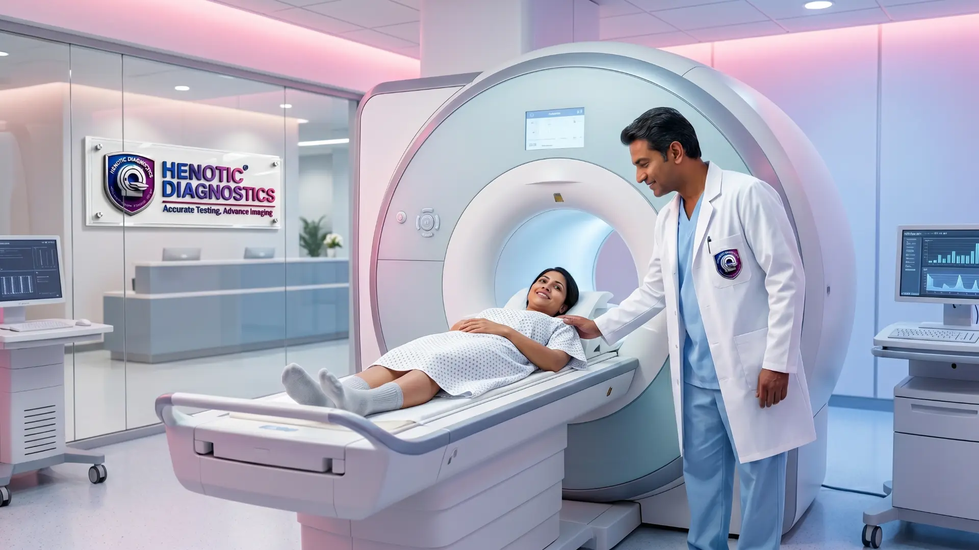
Comprehensive Medical Diagnostic & Radiology Overview
A Radiology Center is a specialized diagnostic facility that utilizes advanced medical imaging technologies—specifically including MRI scan services, CT scan services, PET-CT imaging, ultrasound and sonography, Color Doppler study, and digital mammography—to visualize internal organs, tissue physiology, vascular dynamics, and skeletal structures. Furthermore, Henotic Diagnostics operates a modern Radiology Center equipped with 3.0T wide-bore MRI and 128-slice CT systems, delivering board-certified radiologist interpretations for early disease detection, clinical diagnosis, and therapeutic monitoring.
What Is a Radiology Center & Diagnostic Imaging Suite?
Direct Answer Definition:
Specifically, a Radiology Center is an advanced medical imaging center dedicated to obtaining visual representations of the body’s internal anatomy, organ systems, micro-vascular blood flow, and physiological processes using non-invasive energy forms. Consequently, radiology centers play an essential role in modern healthcare by providing objective anatomical data needed by referring clinicians to diagnose diseases, guide targeted interventional procedures, evaluate traumatic injuries, and monitor therapeutic responses across oncology, neurology, cardiology, orthopedics, and gastroenterology.
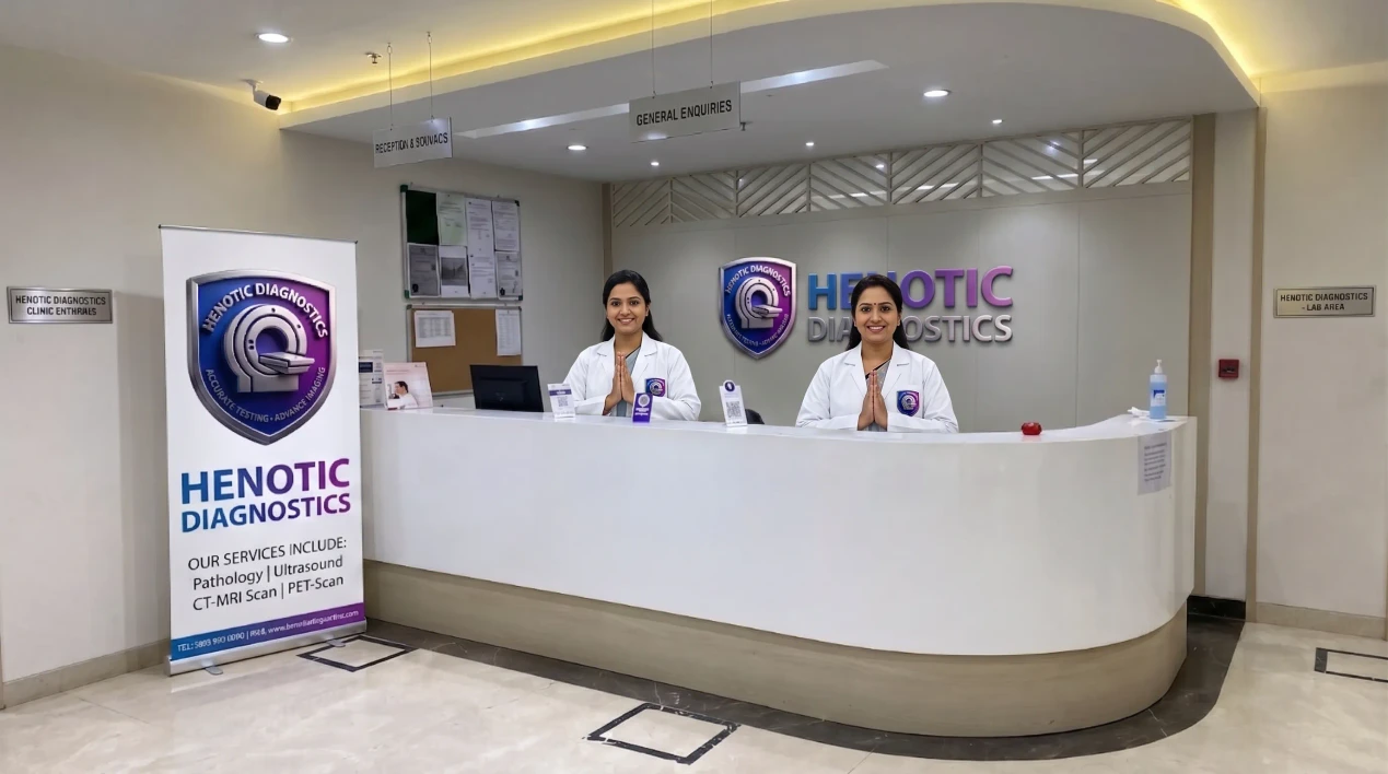
Comprehensive Radiology Center & Medical Imaging Services
1. High-Field 3.0T MRI, 128-Slice CT & Hybrid PET-CT Suite
Specifically, tertiary hospital-grade cross-sectional and molecular oncology diagnostic imaging modalities.
- First, MRI Brain and Epilepsy Protocol
- Second, MRI Spine and Whole Spine Screening
- Third, Joint MRI for Knee, Shoulder, Hip, and Ankle
- Fourth, Abdomen, Pelvis & Prostate MRI
- Fifth, MRCP, MR Angiography & Enterography
- Finally, Cardiac MRI & Whole-Body MRI
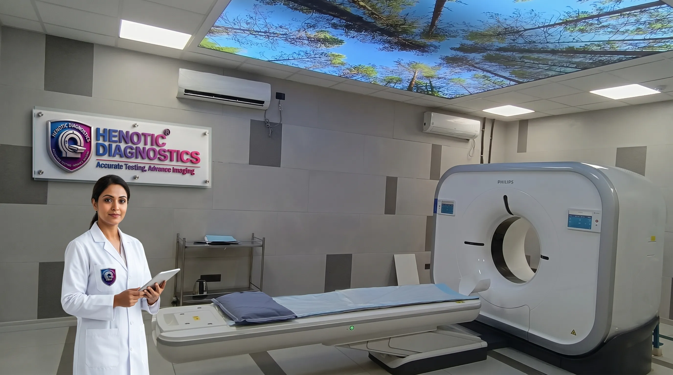
- First, CT Brain & Temporal Bone
- Second, HRCT Chest & Low-Dose Lung CT
- Third, CT Abdomen, Pelvis & CT KUB
- Fourth, CT Coronary & Pulmonary Angiography
- Fifth, CT Enterography & CT Colonography
- Finally, CT-Guided Interventional Procedures
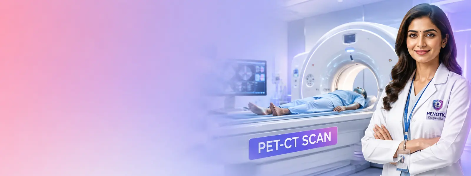
- First, Whole-Body 18F-FDG PET-CT
- Second, PSMA PET-CT (Prostate Oncology)
- Third, DOTA PET-CT (Neuroendocrine)
- Fourth, DOPA PET & FAPI PET Scans
- Fifth, Nuclear Bone Scan & Thyroid Scan
- Finally, Renal Scan (DTPA/EC) & Myocardial Perfusion
2. Acoustic Ultrasound, Color Doppler & Digital Mammography
Additionally, non-ionizing soft-tissue sonography, vascular flow mapping, and low-dose 3D breast screening.
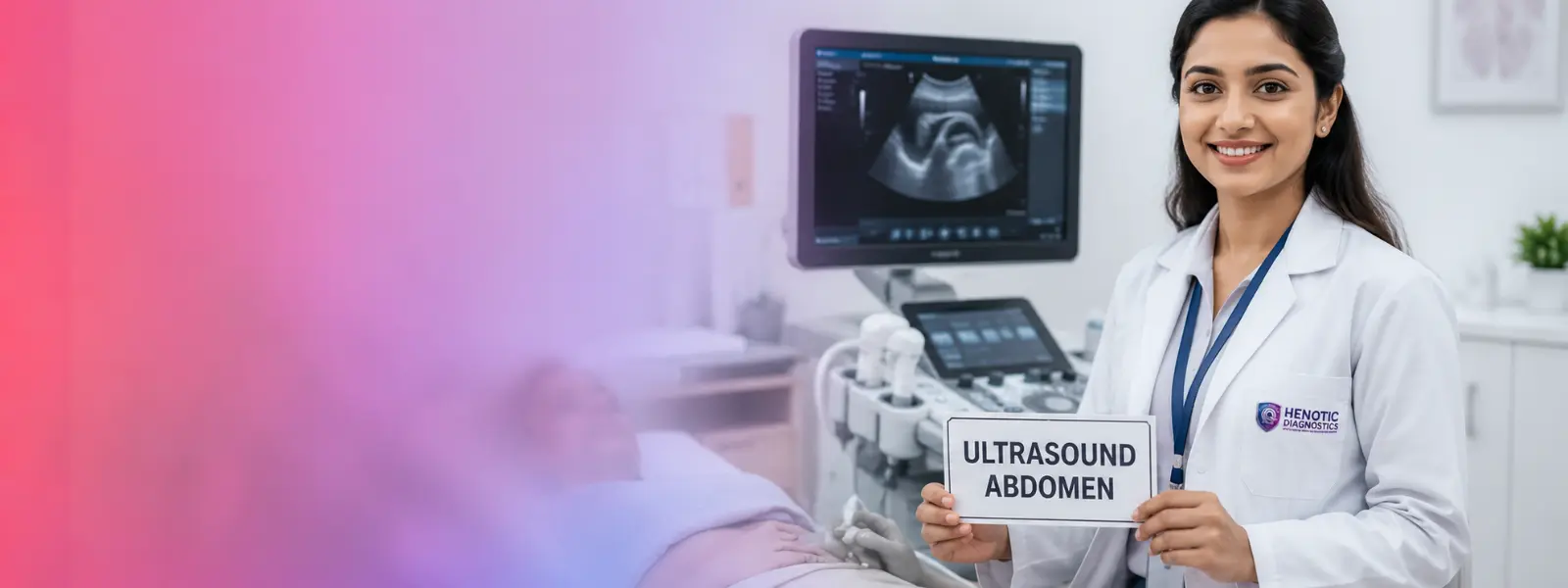
- First, Whole Abdomen & Pelvic Ultrasound
- Second, Ultrasound KUB & Prostate
- Third, Thyroid & Neck Sonography
- Fourth, Musculoskeletal & Soft Tissue USG
- Fifth, Scrotal & Small Parts Ultrasound
- Finally, Transvaginal (TVS) & Transrectal (TRUS)
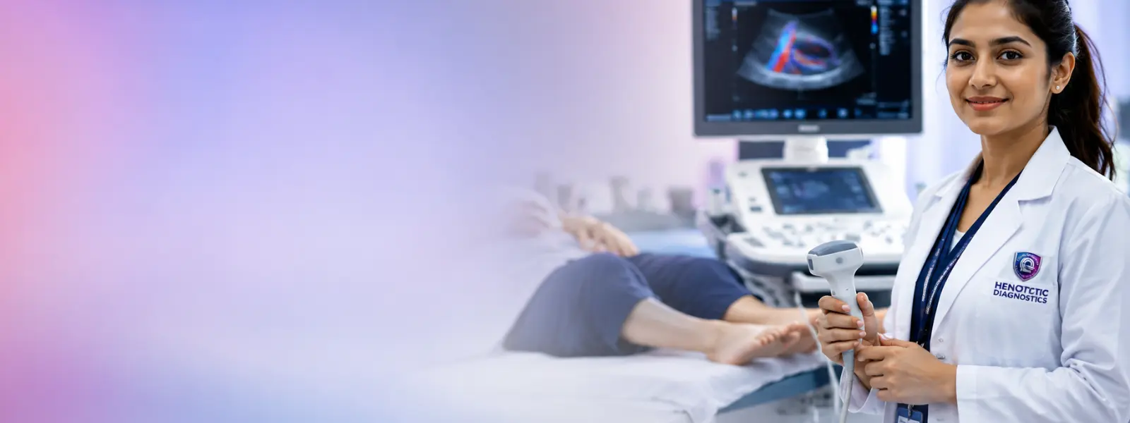
- First, Carotid Artery Doppler
- Second, Arterial & Venous Doppler (Lower/Upper Limb)
- Third, Renal Doppler & Portal Venous Doppler
- Fourth, Obstetric Pregnancy Doppler (Uterine/MCA)
- Finally, Scrotal Varicocele Doppler
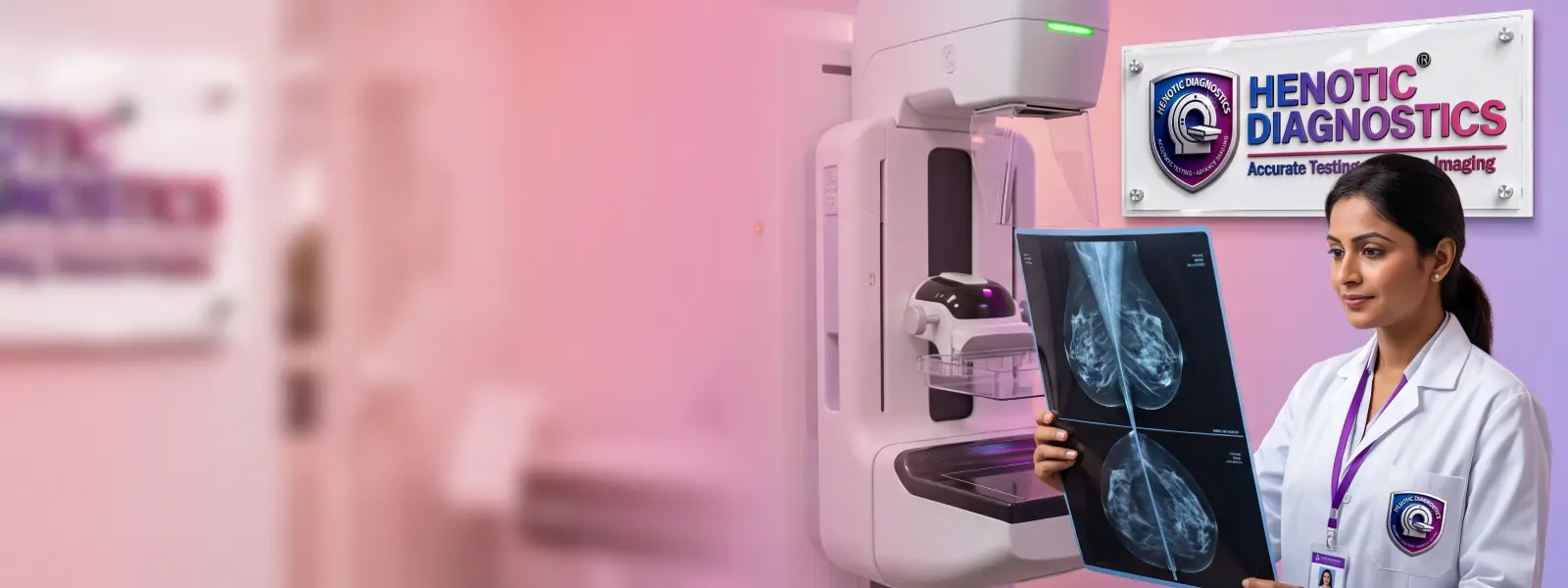
- First, Digital Full-Field Mammography
- Second, 3D Breast Tomosynthesis
- Third, Sonomammography (USG Breast)
- Fourth, Breast MRI & Implants Evaluation
- Finally, Diagnostic Breast Screening Packages
3. Obstetric Fetal Care, Cardiovascular & Interventional Biopsy
Furthermore, PCPNDT-registered fetal growth tracking, 2D echocardiography, and real-time guided biopsies.
- First, Early Pregnancy Viability & Dating Scan
- Second, NT Scan (11-13 Weeks Nuchal Translucency)
- Third, Level 2 Detailed Fetal Anomaly Scan
- Fourth, Fetal Growth Scan & Biophysical Profile
- Finally, Fetal Color Doppler & Echocardiography
- First, CT Coronary Angiography (128-Slice)
- Second, Cardiac MRI & Myocardial Viability
- Third, 2D Echocardiography & Color Doppler
- Fourth, Stress Echocardiography
- Finally, CT Pulmonary Angiography
- First, Ultrasound-Guided FNAC (Fine Needle)
- Second, Ultrasound-Guided Core Biopsy
- Third, CT-Guided Thoracic & Abdominal Biopsy
- Finally, Diagnostic Fluid Aspiration
4. FibroScan Liver Assessment, DEXA Bone Density & Radiography
Moreover, non-invasive liver transient elastography, osteoporosis densitometry, and low-dose digital X-ray.
- First, FibroScan Transient Elastography
- Second, CAP Score Steatosis Quantification
- Finally, Liver Stiffness Measurement (LSM)
- First, DEXA Lumbar Spine & Dual Femur Scan
- Second, Forearm Bone Densitometry
- Finally, Whole-Body Composition Scan
- First, Digital Chest X-Ray (PA/Lateral)
- Second, Musculoskeletal & Joint Radiography
- Third, Spine X-Ray (AP/Lateral/Oblique)
- Fourth, Abdominal X-Ray & KUB
- Fifth, Barium Swallow / Meal Fluoroscopy
- Finally, Hysterosalpingography (HSG) Fluoroscopy
Radiology Center Modality Selection & Clinical Matrix
Neurological, Spine, Joint, Chest & Abdominal Clinical Matrix
| Clinical Area / Indication | Commonly Considered Diagnostic Imaging | Primary Diagnostic Objective |
|---|---|---|
| First, Brain & Neurological Indications | Specifically, MRI Brain examination (3T), CT Brain scan, MR Angiography | Consequently, detect stroke, tumors, aneurysms, demyelination, and acute hemorrhage. |
| Second, Spine & Spinal Cord Indications | Additionally, MRI Spine examination, Whole Spine MRI, CT Spine | Therefore, evaluate disc herniation, spinal stenosis, nerve compression, and trauma. |
| Third, Joints & Soft Tissue Indications | Furthermore, Joint MRI scan, Musculoskeletal Ultrasound | As a result, assess ligament tears, meniscus lesions, tendonitis, and soft-tissue masses. |
| Fourth, Chest & Lungs Indications | For example, HRCT Chest scan, Low-Dose Chest CT, Chest Radiography | Specifically, evaluate interstitial lung disease (ILD), nodules, pneumonia, and fibrosis. |
| Fifth, Abdomen & Viscera Indications | Moreover, Abdominal ultrasound, Abdomen CT scan, MRCP, MRI Abdomen | Consequently, diagnose gallstones, liver pathology, pancreatitis, appendicitis, and renal stones. |
Vascular, Breast, Fetal, Oncology & Bone Density Matrix
| Clinical Area / Indication | Commonly Considered Diagnostic Imaging | Primary Diagnostic Objective |
|---|---|---|
| Sixth, Blood Vessels & Circulation Indications | In addition, Color Doppler study, CT Angiography, MR Angiography | Specifically, detect deep vein thrombosis (DVT), arterial stenosis, varicose veins, and aneurysm. |
| Seventh, Breast Health Indications | Similarly, Digital mammography, Sonomammography, Breast MRI | Therefore, screen microcalcifications, masses, architectural distortion, and dense tissue. |
| Eighth, Pregnancy & Fetal Care Indications | Additionally, Obstetric ultrasound, Level 2 Anomaly Scan, Fetal Doppler | For instance, monitor fetal growth, anatomical development, placental site, and cord flow. |
| Ninth, Cancer Evaluation Indications | Furthermore, PET-CT oncology imaging (FDG/PSMA), Contrast CT, MRI | Consequently, stage primary tumors, detect distant metastases, and evaluate treatment response. |
| Finally, Bone Density Indications | Overall, DEXA bone density scan (Lumbar Spine & Femur) | Specifically, quantify bone mineral density (BMD) and diagnose osteopenia/osteoporosis. |
Why Radiology Center Diagnostic Imaging Quality Matters
Advanced Radiology Center Technology Across Modalities
Digital PACS & Tele-Radiology Consultation Architecture
Zero-Footprint Web DICOM Viewer
First, referring physicians can view high-resolution 3D DICOM cross-sections on desktop or mobile browsers without downloading specialized software.
Instant Patient QR Report Access
Second, patients receive a secure digital portal link and QR code for instant report download and image sharing with consulting specialists.
Encrypted Cloud Data Archiving
Finally, all diagnostic scans are securely archived in redundant cloud storage for long-term comparative evaluation across future hospital admissions.
Online Report Download & Patient Digital Portal Guide
Instant Receipt QR Scanning
First, scan the secure QR code printed on your registration receipt using any smartphone camera to open your personal diagnostic portal dashboard immediately.
Automated WhatsApp & SMS Alerts
Second, as soon as your radiologist signs off your report, a secure encrypted link is sent automatically to your registered WhatsApp and mobile phone.
High-Definition PDF Download
Third, download official digitally signed diagnostic PDF reports containing high-resolution key image slices, quantitative tables, and radiologist impressions.
One-Click Doctor Image Sharing
Finally, generate encrypted temporary viewing links to share full DICOM scan image sets directly with your consulting surgeon or physician.
Radiology Center Services for Referring Doctors & Specialists
Preventive Diagnostic & Radiology Health Checkup Packages
- To begin with, 3.0T Whole-Body Screening MRI
- Additionally, Low-Dose HRCT Chest Scan
- Furthermore, Whole Abdomen & Pelvic Ultrasound
- Moreover, DEXA Lumbar & Dual Femur Density
- Finally, 2D Echocardiography & ECG
- For instance, 128-Slice CT Coronary Angiography
- In addition, Carotid Artery Color Doppler
- Consequently, 2D Echo & Cardiac Perfusion
- As a result, Treadmill Stress Test (TMT)
- Finally, High-Sensitivity Lipid Profile
- Primarily, Digital 3D Breast Tomosynthesis
- Likewise, Sonomammography (Breast USG)
- In addition, Transvaginal Pelvic Sonography (TVS)
- Furthermore, DEXA Bone Mineral Density Scan
- Finally, Thyroid Sonography & Hormonal Panel
- Initially, Whole Spine & Joint MRI Screening
- Also, FibroScan Liver Elastography
- In addition, Ultrasound KUB & Prostate Evaluation
- Consequently, DEXA Fracture Risk Assessment
- Finally, Peripheral Arterial Doppler
Quality Assurance, Phantom Calibration & Peer Double-Reading
How to Prepare for Your Radiology Center Scan
Step-by-Step Patient Diagnostic Experience at Our Radiology Center
Pediatric & Special Support Diagnostic Imaging Protocols
Contrast Imaging Safety at Our Radiology Center
Specifically, contrast media are diagnostic substances administered intravenously, orally, or rectally during a contrast CT scan or contrast MRI scan to temporarily highlight blood vessels, organ borders, and abnormal soft-tissue lesions.
Rapid Emergency Turnaround & Stat Reporting Protocols
Stat Emergency Imaging
First, priority intake protocols ensure rapid scanner access for emergency head CT scans, stroke evaluation, and acute trauma assessment.
Direct Physician Communication
Second, critical or life-threatening radiological findings are communicated directly via phone to referring clinicians by our board-certified radiologist.
Instant Digital PACS Portal
Finally, DICOM image sets and formal signed diagnostic reports are uploaded to our secure PACS cloud portal for instant doctor review.
Radiation Safety Guidelines at Our Radiology Center
Specifically, understanding the distinction between ionizing and non-ionizing imaging technologies helps patients make informed healthcare choices alongside their treating clinicians:
MRI & Ultrasound Safety
Specifically, Magnetic Resonance Imaging uses magnetic fields and radio pulses. Similarly, diagnostic ultrasound uses acoustic sound waves. Consequently, neither modality uses ionizing radiation.
CT & Mammography Safety
Additionally, Computed Tomography and Mammography utilize controlled X-rays. Consequently, modern calibrated scanners follow strict ALARA protocols to minimize dose while preserving image quality.
PET-CT & Nuclear Scans
Furthermore, PET-CT molecular scans and nuclear medicine scans involve tiny amounts of short-lived radiotracers administered to capture metabolic function, decaying rapidly within hours.
Claustrophobia Relief & Patient Comfort Amenities
Senior Reporting Radiologist & Medical Leadership

Dr. Pratibha Patil
Clinical Leadership & Diagnostic Expertise
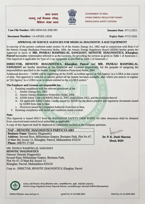
Our Accreditations & Quality Standards
Recognized Statutory Bodies & Accreditation Authorities




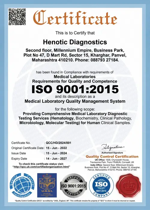
Official Diagnostic Certification Documentation


What Our Patients Say — Patient Testimonials & Clinical Experiences
Frequently Asked Questions About Our Radiology Center
General Radiology Center Services & Diagnostic Modality FAQ
Specifically, core definitions and modality comparisons regarding diagnostic imaging examinations.
Q. What is a radiology center?
Q. What services does a radiology center provide?
Q. Which is better, MRI or CT?
Q. Is MRI safe?
Radiation Safety, MRI Magnetic Safety & PET-CT Oncology FAQ
Additionally, safety guidelines concerning X-ray exposure, magnetic field screening, and tracer uptake.
Q. Does CT use radiation?
Q. Does ultrasound use radiation?
Q. What is a PET-CT scan used for?
Q. What is a Doppler scan?
Q. What is 3T MRI?
Specialized High-Resolution HRCT, Mammography, DEXA & Biopsy FAQ
Furthermore, clinical information on lung scans, 3D breast screening, bone mineral density, and guided tissue sampling.
Q. What is HRCT?
Q. What is mammography?
Q. What is DEXA?
Q. What is FibroScan?
Q. What is an image-guided biopsy?
Corporate Booking Office & Location
