Radiology Services
Because accurate diagnosis leads to better treatment, advanced Radiology Services empower clinical evaluations through high-resolution 3T MRI, multi-slice 128-Slice CT, ultrasound, Doppler, digital mammography, X-ray, PET-CT, DEXA, and nuclear medicine. Additionally, patients can review diagnostic imaging guides on RadiologyInfo.org. Furthermore, emergency findings strictly meet Joint Commission International (JCI) NPSG safety rules. In addition, bone density exams incorporate FRAX® fracture risk scoring. In addition, pediatric scans strictly follow Image Gently® protocols.
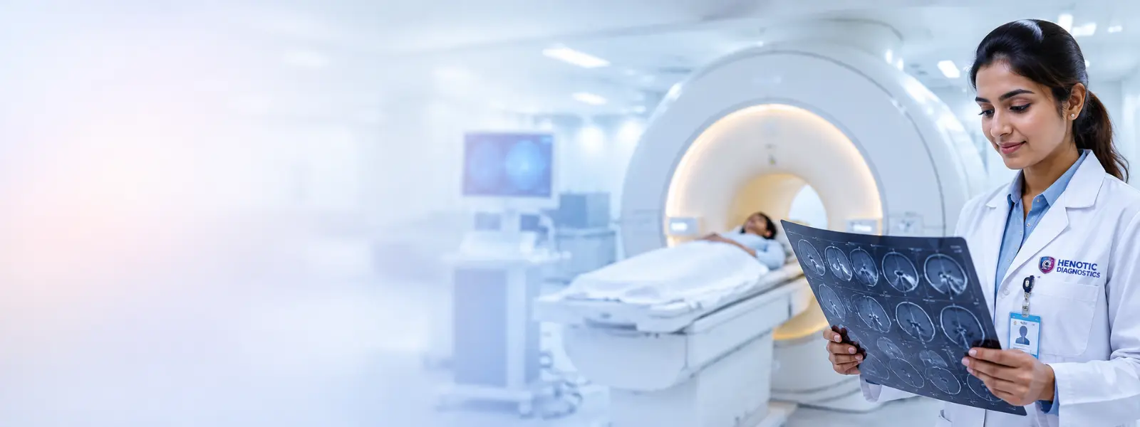
Guided Interventional Radiology & Nuclear Medicine Procedures
As a result, our center provides state-of-the-art minimally invasive image-guided biopsy, aspiration, and nuclear medicine scintigraphy services under real-time guidance.
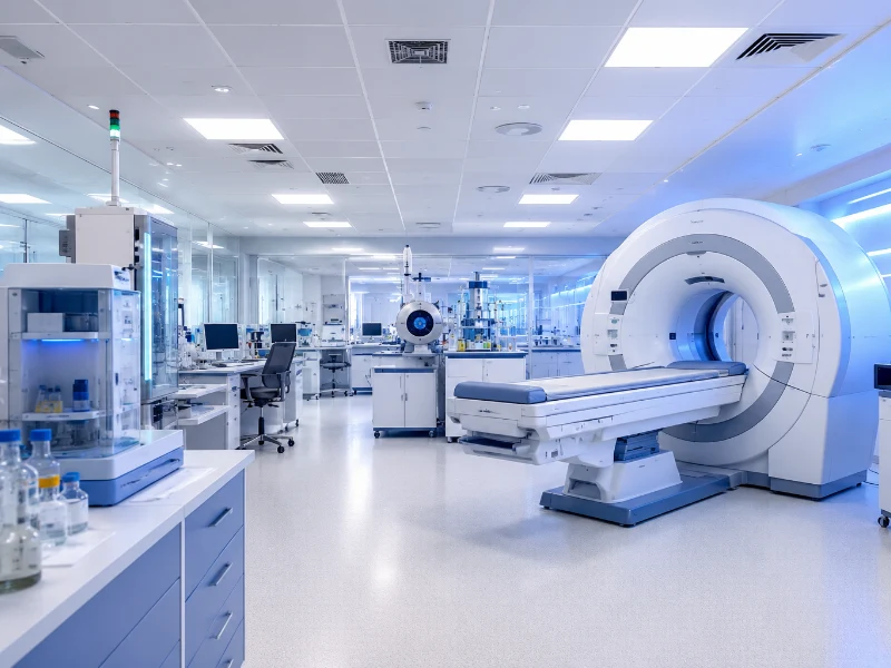


Target Needle Gauge Selection Matrix & Real-Time Tracking
Key Diagnostic Metrics & Trust Signals
How to Share Your DICOM Scan with Consulting Doctors Worldwide
AI-Assisted CADx Triage & Quantitative Imaging Biomarkers in Radiology Services
Emergency Computer-Aided Triage (AI Triage & CADx Engine)
For instance, computer-Aided Detection (CADx) algorithms (AI Triage & CADx Engine) automatically flag acute intracranial hemorrhage, large vessel occlusions (LVO stroke), and acute pulmonary embolism within seconds of DICOM upload, triggering instant radiologist alerts.
Mammographic AI Microcalcification CAD
Notably, concurrent deep learning algorithms analyze full-field digital mammograms, calculating breast tissue density scores and highlighting subtle microcalcification clusters for radiologist verification.
Automated Agatston CAC Calcium Scoring
In fact, deep learning tools automatically measure coronary artery calcium (CAC Agatston Score) on routine non-gated chest CTs, providing opportunistic cardiovascular risk stratification.
AI Radiomics & VCF Spine Fracture CAD
Overall, quantitative radiomic texture analysis evaluates tissue spatial heterogeneity non-invasively, alongside opportunistic deep learning screening for un-reported spinal osteoporotic fractures (VCF) on abdominal/chest CTs.
🫀 AI Aortic Aneurysm & NAFLD Fat Fraction — Radiology Services
As a result, automated volumetric screening measures aortic diameter (>4.0cm expansion alert) and calculates proton density fat fraction (PDFF) for opportunistic NAFLD/MASLD liver steatosis screening.
📊 Quantitative Imaging Biomarkers (QIB) — Radiology Services
Furthermore, ultimately, automated volumetric lesion segmentation, MR proton density fat fraction (PDFF) liver fat quantification, dynamic rCBV perfusion mapping, and CT L3 sarcopenia muscle index calculation.
AI CADx Technical Validation & Performance Metrics
What Are Radiology Services?
In fact, professional Radiology Services represent the core foundation of modern clinical diagnostics, using non-invasive imaging technologies to capture internal anatomical, structural, and functional details of the human body. Notably, by utilizing magnetic fields, sound waves, ionizing radiation, and radiopharmaceuticals, diagnostic imaging enables physicians to accurately identify diseases, assess trauma, monitor organ function, and evaluate treatment efficacy.
In fact, comprehensive Radiology Services play a vital role across all medical specialties, including neurology, cardiology, oncology, orthopedics, gastroenterology, and obstetrics. Furthermore, from detecting microscopic tissue anomalies to guiding precision interventional procedures, specialized imaging ensures evidence-based medical management and enhanced patient safety.
Emergency Critical Value SLA — <30 Minute Direct Physician Notification (International Patient Safety Goals (IPSG))
Because of this, in full compliance with International Patient Safety Goals (IPSG) safety goals, Henotic Diagnostics enforces a mandatory radiologist-to-clinician telephone alert SLA within 30 minutes of scan completion for acute panic findings:
- Specifically, Neuro / Vascular: Acute intracranial hemorrhage, acute cerebral ischemia, aortic dissection, vessel occlusion.
- Thoracic / Abdominal: Specifically, tension pneumothorax, massive pulmonary embolism, bowel perforation, ectopic pregnancy, acute cord compression.
Specifically, 4-Tier Turnaround Time (TAT) SLAs: Routine scans delivered in <12h, Urgent cases in <2h, STAT emergency findings in <30 min, and Critical Callout within <15 min.
Subspecialty Radiology Services Divisions
Neuro-Radiology — Radiology Services
Specifically, comprehensive 3T MRI & CT evaluation for acute stroke, cerebral parenchymal pathology, demyelinating conditions, intracranial MRA, and spinal cord compression.
Musculoskeletal (MSK Radiology) — Radiology Services
Indeed, high-definition multiplanar imaging of meniscus tears, ACL/PCL injuries, rotator cuff tears, cartilage loss, osteomyelitis, and articular joint effusions.
Cardiothoracic Imaging — Radiology Services
Notably, low-dose Moreover, HRCT chest for interstitial lung disease (ILD), ECG-gated CT coronary angiography, pulmonary embolism (CTPA), and cardiac MRI viability imaging.
🌸 Women’s & Breast Imaging — Radiology Services
digital mammography (BI-RADS 0–6), sonomammography, transvaginal pelvic ultrasound, fetal anomaly scanning (Additionally, ISUOG standards), and breast MRI guided biopsy.
🩺 Abdominopelvic Imaging — Radiology Services
In particular, multiparametric prostate MRI (PI-RADS v2.1), MRCP choledocholithiasis mapping, focal liver lesion classification (LI-RADS), and low-dose CT KUB stone protocols.
Pediatric Radiology — Radiology Services
In addition, similarly, radiation-free Fast-MRI protocols (3-minute HASTE scans) and child-sized dose-modulated pediatric sonography following Image Gently® safety standards.
Head & Neck / ENT Imaging Radiology
Specifically, above all, high-resolution sub-millimeter temporal bone CT for conductive hearing loss, paranasal sinus imaging, and high-definition skull base tumor MRI.
🩺 GI & Hepatobiliary Radiology — Radiology Services
In fact, dynamic multi-phase liver CT/MRI, MR Enterography (MRE) for transmural Crohn’s inflammation mapping, and dynamic pelvic floor defecography MRI.
☢️ Nuclear Medicine & Molecular Theranostics
targeted 68Ga-PSMA, 68Ga-DOTATATE, In fact, 18F-FDG PET-CT molecular imaging, I-131 thyroid remnant therapy planning, and whole-body bone scintigraphy.
Advanced Imaging Physics & Standardized Reporting Lexicons
Why Choose Henotic Radiology Services?
Above all, Henotic Diagnostics is committed to delivering clinical precision, patient safety, and compassionate diagnostic care. For this reason, our facility combines high-field scanning technologies with expert radiologist interpretation:
Infrastructure Safety & Radiation Protection Specs
Radiology Services Imaging Modalities
MRI PI-RADS & LI-RADS — Radiology Services
Furthermore, Magnetic Resonance Imaging provides multiplanar, high-resolution soft tissue visualization without ionizing radiation, incorporating PI-RADS v2.1 for prostate assessment and LI-RADS for focal liver lesion evaluation.
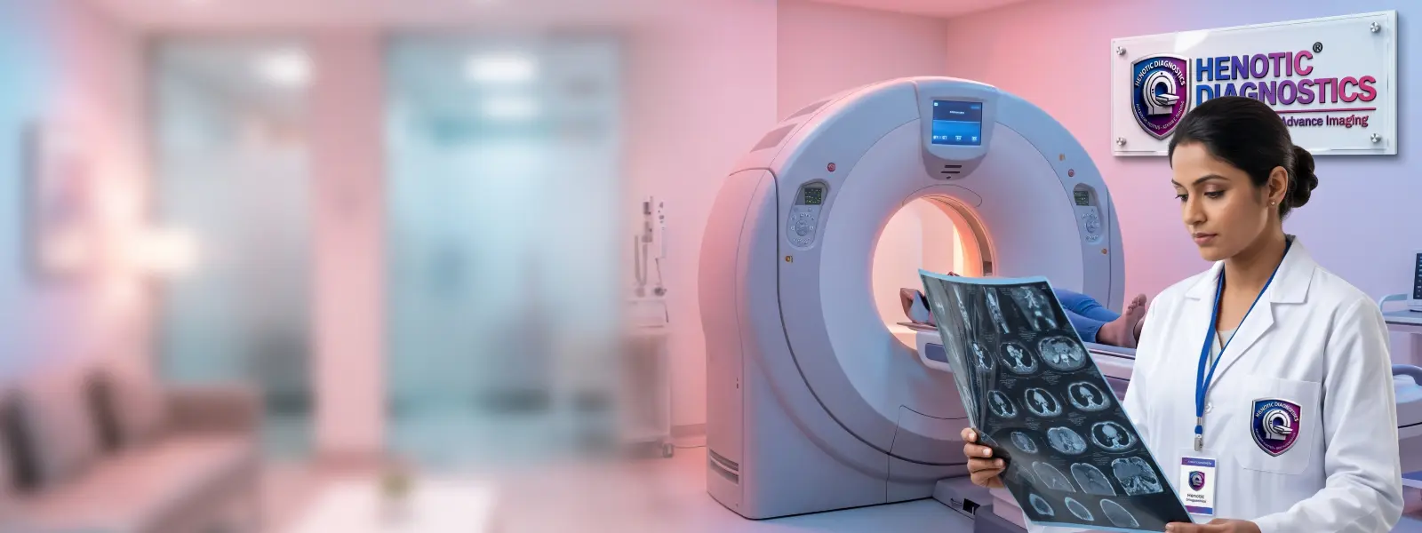
CT Scan 128-Slice MDCT — Radiology Services
Thus, in comparison, Computed Tomography delivers cross-sectional structural imaging of bone, chest, abdominal organs, and vascular networks through rapid multi-slice volumetric acquisition.
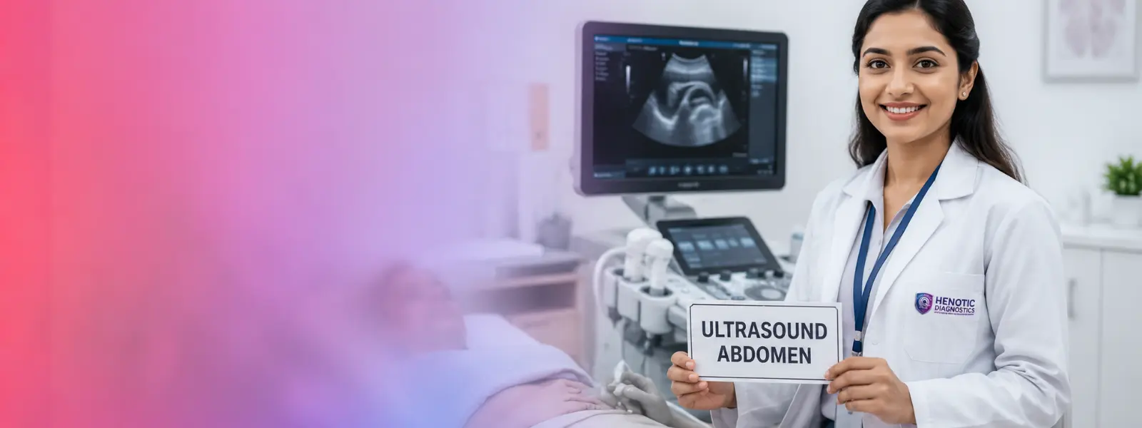
Ultrasound and Sonography TI-RADS — Radiology Services
For this reason, meanwhile, radiation-free imaging utilizes sound waves for real-time visualization of abdominal organs, pelvic structures, soft tissues, thyroid TI-RADS nodule stratification, and obstetric growth monitoring.
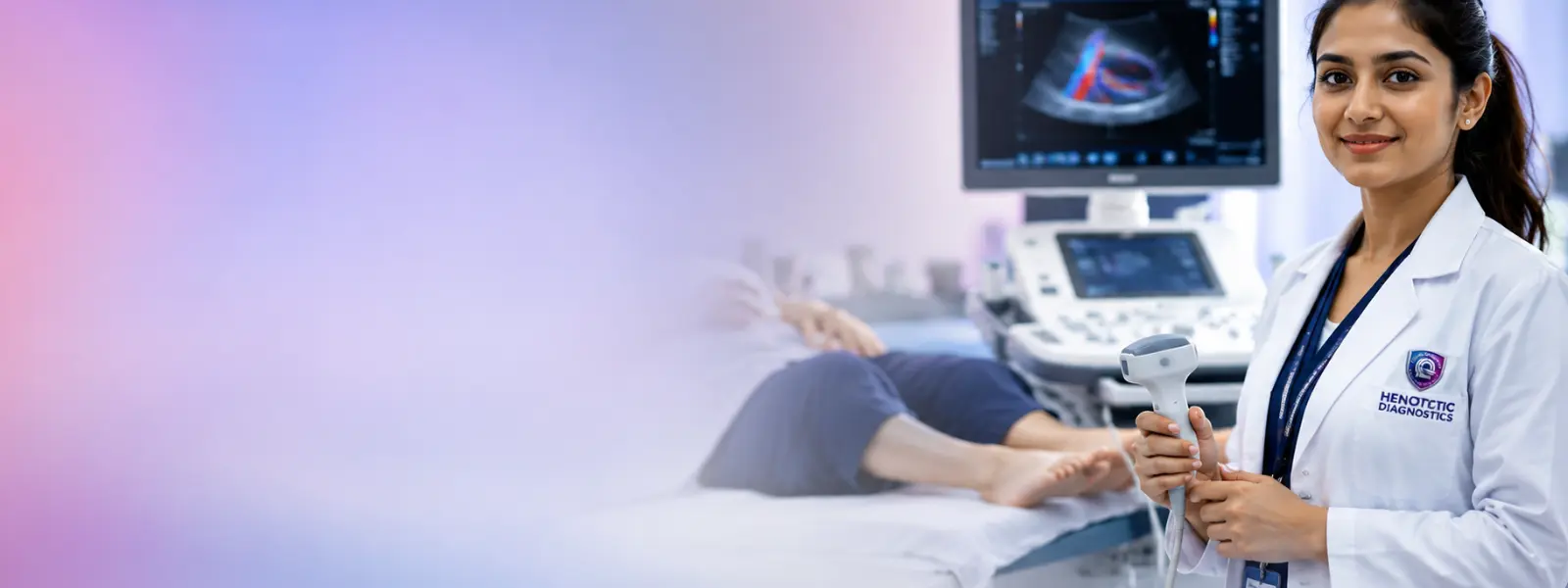
Vascular Color Doppler Radiology Services
In particular, specialized hemodynamic imaging evaluates arterial and venous blood flow velocity, vascular resistance, and hemodynamic perfusion in real time.
Digital Radiography & X-Ray Radiology Services
Accordingly, fundamental digital radiography provides immediate assessment of skeletal integrity, pulmonary conditions, joint alignment, and abdominal emergencies.
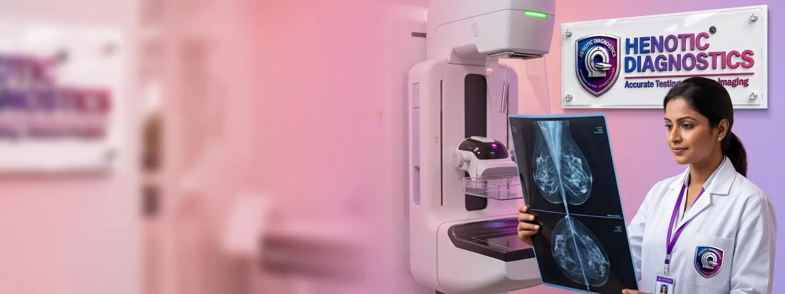
Mammography and Breast Imaging BI-RADS 0–6 — Radiology Services
Specifically, low-dose digital mammography and sonomammography utilize standardized BI-RADS categorization for early detection of microcalcifications and breast tissue irregularities.
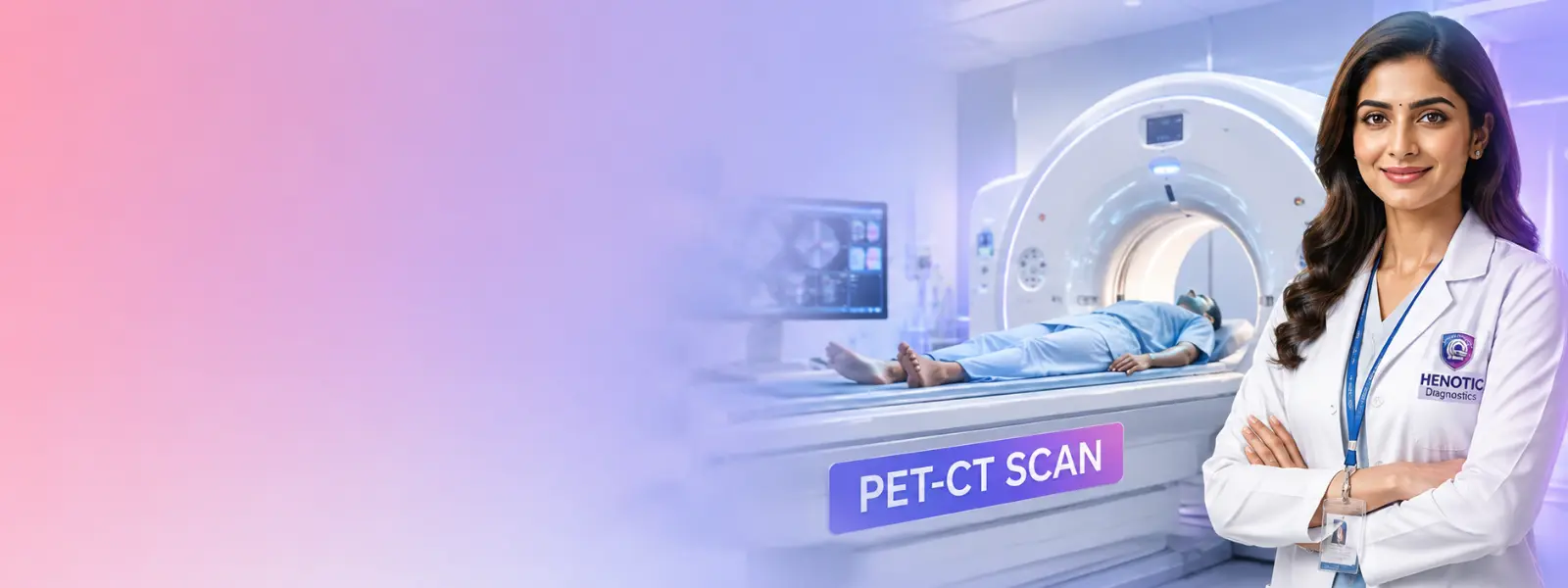
PET-CT RECIST & Theranostics
As a result, integrated Positron Emission Tomography and CT provide metabolic functional insights, employing RECIST 1.1 oncology tracking and Theranostics pairing.
Nuclear Medicine Scintigraphy Radiology Services
Likewise, physiological imaging uses radiotracers to evaluate organ function, bone turnover, thyroid metabolism, and neuroendocrine activity.
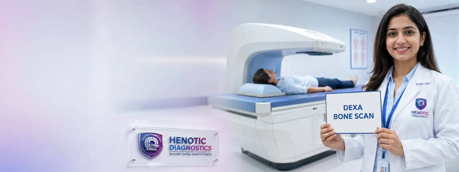
DEXA T-Score / Z-Score — Radiology Services
Additionally, Dual-Energy X-Ray Absorptiometry offers precise quantification of bone mineral density (BMD) for diagnosing osteoporosis and predicting fracture risks.
Advanced Detector Array Physics Across Modalities
Specifically, Interventional Radiology & Image-Guided Procedures
USG / CT Core Needle Biopsy — Radiology Services
High-Precision SamplingAdditionally, real-time image guidance enables automated core tissue sampling of suspicious masses in the breast, liver, lung, kidney, thyroid, retroperitoneum, and subcutaneous soft tissues with minimal discomfort.
Fine Needle Aspiration (FNAC) — Radiology Services
Cytological Micro-SamplingFurthermore, targeted micro-needle cell sampling for rapid cytological evaluation of thyroid nodules (TI-RADS), superficial cervical lymph nodes, salivary gland lesions, and palpable subcutaneous masses.
Fluid Aspiration & Drainage — Radiology Services
Therapeutic ReliefAdditionally, therapeutic ultrasound guidance assists in precise evacuation of symptomatic pleural effusions, abdominal ascites, joint effusions, Baker’s cysts, and intra-abdominal abscesses.
USG-CT Fusion & Hemostatic Patches — Radiology Services
Deep Tissue NavigationMoreover, real-time fusion overlaying ultrasound on CT volumes enables high-precision deep retroperitoneal sampling, followed by bio-collagen hemostatic patch application to prevent post-biopsy hematomas.
Percutaneous Furthermore, Microwave Ablation (MWA / RFA) — Radiology Services
3D Cone-Beam CT GuidedAs a result, percutaneous microwave (MWA) and radiofrequency (RFA) thermal tumor ablation for liver, lung, and renal lesions incorporating intra-procedural 3D Cone-Beam CT trajectory planning.
PTBD Drainage & NGS Biopsy — Radiology Services
Genomic Sequencing AdequacyAdditionally, fluoroscopic PTBD relieves obstructive jaundice, while high-yield core biopsy protocols guarantee specimen adequacy for Next-Generation Sequencing (NGS) target mutations (EGFR/ALK/PD-L1).
🚗 Post-Biopsy Companion Escort Policy & Coagulation Thresholds
Interventional Complication Audit & Thermal Ablation Parameters
Interventional Radiology Pre-Procedure Hemostasis & Anticoagulation Hold Thresholds
| Coagulation / Lab Parameter | SIR / CIRSE Bleeding Risk | Required Lab Threshold | Medication Hold SLA |
|---|---|---|---|
| Accordingly, Superficial FNAC / Biopsy | First, category 1 (Low Risk) | Finally, INR ≤ 1.8, Platelets ≥ 50,000 / μL | In particular, aspirin: Continue | LMWH: Hold 12h prophylactic |
| As a result, Deep Organ Biopsy / PTBD / Ablation | Meanwhile, category 2 (High Risk) | Thus, INR ≤ 1.5, Platelets ≥ 50,000 / μL | Hence, warfarin: Hold 5 days (INR < 1.5) | DOACs: Hold 48h |
| Additionally, Low Molecular Weight Heparin (LMWH) | Indeed, anticoagulant SLA | In light of this, anti-Xa activity baseline | With this in mind, hold 24h (therapeutic) / 12h (prophylactic) |
| Specifically, Unfractionated Heparin (UFH) | Furthermore, anticoagulant SLA | In addition, normal aPTT reference | Consequently, hold IV infusion 4–6 hours prior to procedure |
| Prasugrel & Ticagrelor | As a result, antiplatelet SLA | For instance, platelets ≥ 50,000 / μL | In fact, prasugrel: Hold 7–10 days | Ticagrelor: Hold 5 days |
| Notably, NSAIDs (Ibuprofen / Naproxen) | Moreover, NSAID Protocol | In contrast, normal baseline clearance | Likewise, hold 1–3 days depending on agent half-life |
Post-Procedure Recovery & Complication Management SLA — Radiology Services
In addition, post-biopsy complication management protocols require monitored observation (2 hours for superficial FNAC; 4 hours with post-procedure erect expiratory chest X-ray for lung biopsy pneumothorax exclusion), bio-collagen hemostatic patch application, and compression or thrombin injection for hematomas.
MRI Radiology Services
Brain MRI Radiology Scans
As a result, this scan provides detailed evaluation of cerebral parenchyma, stroke identification, demyelinating conditions, cranial nerve pathology, and intracranial lesions.
Spine MRI Radiology Services
Consequently, it offers high-definition visualization of cervical, thoracic, and lumbar intervertebral discs, spinal cord signal intensity, and nerve root compression.
MRI Knee & Joint
Therefore, it delivers comprehensive soft-tissue diagnosis for meniscus tears, ACL/PCL ligament injuries, tendonitis, articular cartilage loss, and joint effusions.
MRI Shoulder & Thigh
However, detailed multiplanar imaging accurately evaluates rotator cuff tears, labral lesions, shoulder impingement syndrome, and soft tissue masses.
Abdominal MRI & Pelvis MRI LI-RADS & mpMRI
In addition, multiparametric prostate MRI (mpMRI) incorporates ultra-high diffusion (b=2000 s/mm²) for detecting clinically significant cancer (>Gleason 3+4), while DCE MRI assesses focal liver lesions (LI-RADS), endometriosis, and silicone breast implant rupture integrity.
MRCP (Cholangiopancreatography)
As a result, non-invasive fluid-sensitive evaluation maps intrahepatic biliary ducts, common bile duct stones (choledocholithiasis), and pancreatic ducts.
MR Angiography (MRA)
Likewise, radiation-free contrast and non-contrast (Time-of-Flight) mapping evaluates intracranial cerebral arteries, carotid bifurcations, and renal arteries.
Diffusion Tensor Imaging (DTI) & DSC MR Perfusion — Radiology Services
Specifically, DTI white matter tractography maps brain neural pathways for surgical planning, while Dynamic Susceptibility Contrast (DSC) MR Perfusion quantifies cerebral blood volume (rCBV) in stroke penumbra.
MR Enterography (MRE) & Pelvic Defecography MRI — Radiology Services
In contrast, oral iso-osmolar polyethylene glycol contrast protocols deliver high-sensitivity non-ionizing transmural Crohn’s inflammatory bowel disease evaluation and dynamic pelvic floor organ prolapse assessment.
Whole-Body MRI (MY-RADS) & Eovist Liver MRI — Radiology Services
For example, radiation-free Whole-Body MRI adhering to MY-RADS guidelines evaluates multiple myeloma, alongside Eovist (Gadoxetic Acid) dynamic hepatobiliary imaging differentiating FNH from hepatocellular adenoma.
Specialized Neuro & Cardiac MRI
Finally, advanced protocols include Diffusion-Weighted Imaging (DWI), MR Spectroscopy (MRS), functional MRI (fMRI), 4D Flow cardiac velocity mapping, and myocardial viability assessment.
Anti-Claustrophobia Patient Comfort Toolkit
1. Noise-Canceling Music Headphones
Notably, high-fidelity pneumatic headphones playing your choice of relaxing music playlists, radio channels, or acoustic soundscapes to mute gradient noise.
2. Emergency Hand Squeeze Ball
Specifically, hold a soft pneumatic squeeze ball to communicate instantly with your technologist at any second for immediate voice assistance.
3. 70cm Bore & Open-Sky Prisms
In particular, extra-wide 70cm scanner aperture with specialized angled mirror prisms providing a panoramic view out of the bore toward the room window.
4. Family Companion Protocol
Because of this, a screened family member or friend is welcome to sit directly beside you inside the MRI room holding your hand during scanning.
5. Hydraulic Low-Entry Table
Ultimately, hydraulic ultra-low scanner table height (48cm clearance) for effortless transfer from wheelchairs with memory foam joint cushions.
In-Bore 4-7-8 Rhythmic Breathing Anti-Anxiety Technique:
Pulse Sequence Parameters & RF Coil Specs
CT Scan Radiology Services
CT Brain
In addition, initially, this test provides rapid cross-sectional evaluation for acute intracranial hemorrhage, ischemic infarction, traumatic skull fractures, and cerebral edema.
HRCT Chest & Low-Dose CT Screening LUNG-RADS 1.1
Consequently, high-Resolution Computed Tomography (HRCT) evaluates interstitial lung disease (ILD) and pulmonary fibrosis, incorporating LUNG-RADS 1.1 risk stratification for pulmonary nodules ≥ 6mm.
CT Abdomen & Pelvis
In fact, multi-phase contrast imaging assesses acute appendicitis, diverticulitis, bowel obstruction, liver metastases, and abdominal trauma.
CT KUB & Dual-Energy Spectral CT (DECT)
Overall, low-dose volumetric protocols provide high sensitivity for renal calculi. Likewise, Dual-Energy Spectral CT (DECT) enables gout uric acid crystal mapping, virtual monoenergetic imaging, and tissue iodine quantification.
Angiography CT Radiology Services
Ultimately, 3D arterial vascular mapping evaluates thoracic/abdominal aortic aneurysms, pulmonary embolism (CTPA), and peripheral vascular stenosis.
CT Coronary Angiography CAD-RADS 2.0
Ultimately, ECG-gated non-invasive visualization detects coronary artery lumen narrowing, atherosclerotic plaque composition, and Calcium Scoring, codified under CAD-RADS 2.0 (Grades 0–5, with V vulnerable plaque and P2 main vessel modifiers).
ECG-Gated TAVR / TAVI Structural Heart CT — Radiology Services
Hence, specialized structural heart protocols provide sub-millimeter aortic root annulus geometry sizing, coronary ostia height measurement, and peripheral vascular access evaluation.
CT Colonography (Virtual Colonoscopy – C-RADS)
In particular, low-dose volumetric virtual colonoscopy provides non-invasive colorectal polyp screening adhering to C-RADS criteria, alongside 3D CT virtual airway bronchoscopy.
CT Fractional Flow Reserve (FFR-CT) & Cochlear CT — Radiology Services
Accordingly, computational fluid dynamics evaluate the hemodynamic severity of coronary stenoses (FFR-CT <= 0.80), alongside sub-millimeter temporal bone CT for cochlear implant planning.
CT Enterography
Specifically, multi-detector CT Enterography utilizes neutral oral contrast to evaluate small bowel Crohn’s disease, mural thickening, and mucosal inflammation.
What to Expect During IV Contrast CT Scans — Radiology Imaging
ACR Contrast Manual v2023 Safety Protocols — Radiology Services
X-Ray Tube Geometry & Spectral Spectral Accuracy
Ultrasound Radiology Services
Abdominal & Whole Abdomen Ultrasound
Overall, it ensures thorough evaluation of liver echogenicity, gallstones (cholelithiasis), splenomegaly, pancreatic structure, and renal parenchymal thickness.
Pelvic Ultrasound O-RADS 1–5
Therefore, transabdominal and transvaginal sonography evaluates uterine leiomyomas, endometrial thickness, and adnexal mass malignancy risks using O-RADS 1–5 criteria.
Kidney & Urinary Tract Ultrasound
Additionally, targeted renal assessment identifies hydronephrosis, renal cysts, bladder wall thickening, and post-void residual urine volume.
Thyroid Ultrasound TI-RADS
Furthermore, high-resolution sonography offers standardized TI-RADS nodule risk stratification, parathyroid adenomas, and cervical lymph node evaluation.
Breast Ultrasound (Sonomammography) BI-RADS
As a result, targeted acoustic assessment differentiates solid breast masses from fluid-filled benign cysts using BI-RADS reporting standards.
Obstetric Ultrasound
Likewise, it provides comprehensive fetal gestational tracking, nuchal translucency (NT) screening, anomaly scans, and fetal biometry.
Soft Tissue & Musculoskeletal Ultrasound
Finally, dynamic acoustic imaging evaluates superficial lipomas, tendon tears, joint effusions, and subcutaneous abscesses.
Liver Shear Wave Elastography (SWE / FibroScan) — Radiology Services
Moreover, non-invasive acoustic shear wave elastography measures liver tissue stiffness (in kPa), providing objective METAVIR fibrosis staging (F0 to F4) for chronic liver disease.
Indeed, Contrast-Enhanced Ultrasound (CEUS) & SMI Microvascular Flow — Radiology Services
second-generation microbubble acoustic contrast (sulfur hexafluoride) delivers non-nephrotoxic focal liver lesion characterization in renal failure patients, alongside Superb Microvascular Imaging (SMI) for low-velocity microvessel detection.
22 MHz MSK Micro-Ultrasound & TCD Vasospasm — Radiology Services
In addition, ultra-high frequency 22 MHz transducers evaluate peripheral nerve entrapment (carpal tunnel area >10mm²), alongside Transcranial Doppler (TCD) cerebral vasospasm monitoring (>120 cm/s).
Matrix Transducer Physics & Acoustic Safety Parameters
Doppler Ultrasound Radiology Services
Carotid Doppler Ultrasound Scan
First, assessment of carotid artery intima-media thickness (CIMT) and atheromatous plaque burden helps stratify stroke risk.
Lower Limb Doppler Study
Additionally, second, arterial and venous flow evaluation detects Deep Vein Thrombosis (DVT), chronic venous insufficiency, and peripheral arterial occlusion.
Upper Limb Doppler Scan
Furthermore, third, vascular flow assessment evaluates subclavian steal syndrome, thoracic outlet arterial compression, and upper extremity thrombosis.
Peripheral Arterial Doppler Study
As a result, hemodynamic Doppler waveform analysis and Ankle-Brachial Index (ABI) correlation assess peripheral artery disease (PAD).
Middle Cerebral Artery Doppler Study
Consequently, transcranial Doppler imaging measures fetal MCA peak systolic velocity to detect fetal anemia and intracranial hemodynamics.
Renal Doppler Scan
Therefore, renal artery flow velocity measurement and resistive index (RI) calculation diagnose renovascular hypertension.
Umbilical Artery Doppler Study & Uterine Artery Doppler Study
However, placental resistance Doppler monitoring evaluates intrauterine growth restriction (IUGR) and pre-eclampsia risk.
Vascular Color Doppler Study
In addition, ultimately, comprehensive systemic color Doppler mapping evaluates vascular malformations, varicose veins, and aneurysm flow patterns.
Penile Color Doppler with Pharmacological Injection (ICI)
In contrast, real-time arterial inflow (PSV > 35 cm/s) and venous leak resistance assessment evaluate vasculogenic erectile dysfunction.
Venous Doppler
In fact, non-invasive Venous Doppler sonography evaluates lower and upper extremity venous blood flow patterns to detect venous incompetence.
Venous Insufficiency Scan
Likewise, specialized Venous Insufficiency Scan protocols quantify saphenofemoral junction valvular reflux and superficial vein dysfunction.
Deep Vein Thrombosis Scan
Overall, rapid Deep Vein Thrombosis Scan examination performs compression ultrasonography to identify occlusive venous thrombi quickly.
Hemodynamic Doppler Formulas & Angle Calibration
X-Ray Radiology Services
Digital Chest & Dual-Energy Subtraction Radiography (DES) — Radiology Services
For example, standard PA/lateral projections and Dual-Energy Subtraction Radiography (DES) separate bone from soft tissue, unmasking calcified pulmonary nodules hidden behind ribs.
Skeletal Radiography — Radiology Services
For instance, high-resolution bone imaging assesses acute cortical fractures, joint dislocations, bone tumors, and osteomyelitis.
Spinal Radiography & Full-Length EOS Standing Radiography — Radiology Services
Notably, cervical, thoracic, and full-length EOS standing radiography series utilize automated digital stitching software for precise scoliosis Cobb angle measurement and leg length discrepancy calculation.
Abdominal X-Ray (KUB) — Radiology Services
In fact, plain film imaging checks bowel gas patterns, intestinal perforation (free air under diaphragm), and radio-opaque urinary stones.
Contrast Fluoroscopy / HSG — Radiology Services
Finally, real-time dynamic imaging procedures include Hysterosalpingography (HSG) for fallopian tube patency and Barium swallow studies.
Flat Panel Detector (FPD) CsI Technical Specs
Mammography and Breast Radiology Services
BI-RADS Classification Standard — Radiology Services
Full-Field Digital Mammography System with 3D Tomosynthesis BI-RADS Category 1–2
As a result, low-dose 3D digital breast tomosynthesis captures 1mm thin tissue slices, eliminating overlapping tissue confusion across American College of Radiology (ACR) Density Categories A, B, C, and D.
Diagnostic Mammography Category BI-RADS 3–5
In contrast, targeted compression and spot-magnification views investigate palpable breast masses, localized pain, or skin changes.
Breast Ultrasound (Sonomammography)
Notably, complementary acoustic evaluation characterizes solid vs. Additionally, cystic breast lesions, especially in mammographically dense tissue.
Breast MRI & Guided Biopsy ACR BI-RADS 6
Specifically, ultimately, high-contrast multiparametric breast imaging enables high-risk screening, pre-operative staging, and image-guided tissue sampling.
3D Mammography
Furthermore, advanced 3D Mammography captures three-dimensional volumetric images of dense breast tissue to detect occult lesions early.
Breast Imaging
Specifically, comprehensive Breast Imaging integrates multimodality screening and diagnostic protocols to ensure accurate structural evaluation.
3D Breast Imaging
Consequently, state-of-the-art 3D Breast Imaging delivers high-resolution slice projections to minimize tissue overlap false positives.
Breast Screening
In addition, routine Breast Screening facilitates proactive asymptomatic health monitoring to identify subtle microcalcifications promptly.
Breast Cancer Screening
As a result, specialized Breast Cancer Screening utilizes low-dose digital mammography standards for early oncological detection.
Breast Diagnostics
Moreover, targeted Breast Diagnostics evaluate suspicious palpable masses and focal asymmetrical densities with clinical precision.
Digital Breast Tomosynthesis
Therefore, high-definition Digital Breast Tomosynthesis acquires low-dose cross-sectional sweeps for superior diagnostic confidence.
Patient Comfort & Pain Mitigation Mammogram Guide
- For example, Curved Compression Paddles: Flexible anatomical paddles adjust to your natural body contours, minimizing pressure discomfort.
- Best Scheduling Timing: Schedule your mammogram 7 to 10 days after your menstrual cycle when breast tissue is least sensitive.
- Additionally, 100% Female Technologists: All breast scans are conducted with gentle care by certified, empathetic female mammographers.
- In contrast, Deodorant & Powder Preparation Notice: Please do not apply underarm deodorants, talcum powders, or body lotions on the morning of your scan, as metallic aluminum microparticles can mimic microcalcifications on mammograms.
- Silicone / Saline Breast Implants: Our mammographers use specialized Eklund implant displacement technique views to ensure safe, comprehensive imaging of native breast tissue without implant distortion.
Contrast-Enhanced Mammography & Dose Limits
PET-CT Scans, Theranostics & RECIST 1.1 Oncology Tracking in Radiology Services
Oncology RECIST 1.1 Radiology Services Frameworks
Ultimately, at Henotic Diagnostics, consultant radiologists evaluate oncological scan series using standardized quantitative response assessment criteria to deliver objective treatment efficacy metrics for treating oncologists and tumor boards:
Standardized RECIST 1.1 (Solid Tumor Anatomical Criteria)
First, measures sum of longest diameters (SLD) of target lesions: Complete Response (CR) (disappearance of all lesions), Partial Response (PR) (≥30% decrease in SLD), Progressive Disease (PD) (≥20% increase in SLD or new lesions), and Stable Disease (SD).
RECIST 1.1 • Unidimensional MeasurementPERCIST 1.0 (Metabolic SUVpeak Response)
For instance, tracks tumor SULpeak (SUV corrected for lean body mass): Complete Metabolic Response (CMR), Partial Metabolic Response (PMR) (≥30% SULpeak decrease), Progressive Metabolic Disease (PMD) (≥30% SULpeak increase), and Stable Metabolic Disease (SMD).
PERCIST 1.0 • SULpeak Metabolic MetricLugano 2014 & Deauville 5-Point Scale
In fact, standardized lymphoma response classification comparing 18F-FDG uptake against mediastinal blood pool and liver SUV standards: Deauville Scores 1 to 5 (Score 1-3 = Complete Response; Score 4-5 = Residual Metabolic Disease).
Lugano 2014 • Deauville 1–5 ScaleiRECIST (Immuno-Oncology Response)
Indeed, specialized criteria for patients receiving immune checkpoint inhibitors (PD-1/PD-L1 inhibitors), differentiating early immune cell infiltration flare (pseudo-progression) from unconfirmed progressive disease (iUPD) vs. Notably, confirmed progression (iCPD).
iRECIST • Immunotherapy Pseudo-ProgressionTheranostic Radiology Services Radionuclide Matrix
Radionuclide Dosage & Target Indication Specifications
| Cancer Indication | Diagnostic PET/Scintigraphy Tracer | Therapeutic Radionuclide Partner | Target Mechanism & Action |
|---|---|---|---|
| Similarly, metastatic Prostate Cancer (mCRPC) | Overall, 68Ga-PSMA-11 / 18F-DCFPyL | 177Lu-PSMA-617 (Pluvicto) | Ultimately, binds Prostate-Specific Membrane Antigen (PSMA) for targeted beta-particle tumor cell destruction. |
| neuroendocrine Tumors (GEP-NETs) | For this reason, 68Ga-DOTATATE / DOTANOC | Accordingly, 177Lu-DOTATATE (Lutathera) | In particular, targets somatostatin receptor subtype 2 (SSTR2) for Peptide Receptor Radionuclide Therapy (PRRT). |
| Differentiated Thyroid Cancer (DTC) | Finally, 123I / 99mTc-Pertechnetate / 18F-FDG | In particular, 131I (Radioactive Iodine NaI) | Similarly, sodium-Iodide Symporter (NIS) targeting for thyroid remnant ablation and metastatic disease eradication. |
| pheochromocytoma & Neuroblastoma | Thus, 18F-DOPA / 123I-MIBG | 131I-MIBG (Azedra) | Additionally, norepinephrine transporter system targeting for high-activity chromaffin tumor radionuclide therapy. |
| Indeed, Castrate-Resistant Bone Metastases | In light of this, 99mTc-MDP / 18F-NaF Bone PET | With this in mind, 223Ra-Dichloride (Radium-223 Xofigo) | Furthermore, alpha-emitting calcium mimetic target destroying osseous metastases while sparing adjacent bone marrow. |
| Specifically, unresectable Liver Tumors (HCC / Mets) | Furthermore, 99mTc-MAA Hepatic Perfusion SPECT/CT | 90Y-Microspheres (TARE / SIRT) | Thus, hepatic arterial embolization planning for Selective Internal Radiation Therapy (SIRT). |
PET-CT Radiology Services Core Examinations
18F-FDG Whole Body PET-CT RECIST 1.1 & Deauville
Notably, similarly, metabolic glucose uptake mapping enables early primary staging, restaging, and treatment monitoring across lung, colorectal, breast, lymphoma, and head & neck cancers.
PSMA PET-CT Scan (68Ga-PSMA-11 & 18F-DCFPyL) Theranostics
Additionally, above all, ultra-high sensitivity PSMA receptor imaging detects biochemical prostate cancer recurrence at PSA levels as low as 0.2 ng/mL, guiding 177Lu-PSMA theranostic selection.
DOTA PET-CT Scan (68Ga-DOTATATE / DOTANOC) PRRT Planning
In fact, somatostatin receptor subtype 2 (SSTR2) PET imaging offers high-sensitivity detection of gastroenteropancreatic NETs, carcinoids, and 177Lu-DOTATATE PRRT candidate screening.
DOPA PET-CT Diagnostic Scan (18F-DOPA) Neuroendocrine
Overall, specialized amino acid transporter PET evaluation analyzes medullary thyroid carcinoma (MTC), pheochromocytomas, paragangliomas, and congenital hyperinsulinism.
68Ga-FAPI & 18F-FES (Fluoroestradiol) PET-CT Radiomics
Overall, targeted Fibroblast Activation Protein Inhibitor (FAPI) and 18F-FES estrogen receptor PET imaging map stroma-rich epithelial tumors and ER+ metastatic breast cancer.
18F-FET Glioma & 18F-Florbetapir Brain PET-CT Neuro-Oncology
For this reason, hence, quantitative 18F-FET amino acid PET differentiates recurrent brain tumor from radiation necrosis, alongside 18F-Florbetapir amyloid plaque imaging for Alzheimer’s diagnosis.
18F-FDG Cardiac Viability & Sarcoidosis PET-CT Cardiology
Thus, combined myocardial perfusion/glucose metabolic imaging identifies hibernating viable myocardium prior to revascularization, alongside cardiac sarcoidosis inflammatory mapping.
Quantitative Imaging Biomarkers (QIB) in PET-CT
- Accordingly, SUVmax & SUVpeak: Standardized Uptake Value peak concentration measuring tumor metabolic aggressiveness.
- Likewise, Metabolic Tumor Volume (MTV cm³): Volumetric quantification of active metabolizing tumor tissue.
- Besides this, Total Lesion Glycolysis (TLG = SUVmean × MTV): Integrated product representing total metabolic tumor burden.
PET-CT Scan Day Patient Experience Walkthrough
1. 24-Hour Low-Carb Pre-Scan Diet — Radiology Services
Furthermore, ultimately, 24 hours before scan: Eat high-protein, zero-sugar foods (avoid rice, sugar, bread, alcohol, and strenuous exercise), followed by 6 hours of fasting.
2. Arrival & Glucose Check — Radiology Services
Notably, check-in and quick finger-prick blood glucose test (<150 mg/dL required for optimal 18F-FDG scan clarity).
3. 60-Minute Recliner Rest — Radiology Services
First, relax comfortably in a private heated recliner chair while the radiotracer circulates peacefully.
4. 15-Minute Scan — Radiology Services
Hence, lie on the padded scanner couch for a brief 15 to 20 minute whole-body scan acquisition.
5. Post-Scan Hydration — Radiology Services
In particular, drink plain water after your scan to help flush out the radiotracer naturally within a few hours.
Time-of-Flight (TOF) Molecular Physics
DEXA and Bone Density Radiology Services
Lumbar Spine BMD DEXA T-Score ≥ -1.0
Accordingly, quantification of bone density across L1-L4 vertebrae evaluates axial skeleton fracture risk.
Dual Femur / Hip DEXA Osteopenia / Osteoporosis
Specifically, precision measurement of femoral neck and total hip mineral density diagnoses osteoporosis accurately.
Vertebral Fracture Assessment (VFA) & TBS Micro-Architecture — Radiology Services
In addition, ultimately, low-dose lateral spine DEXA Vertebral Fracture Assessment (VFA) detects occult vertebral height loss, incorporating Trabecular Bone Score (TBS) micro-architecture, WHO FRAX® fracture risk, and Visceral Adipose Tissue (VAT cm²) metabolic area quantification.
Phantom QA Calibration & LSC Precision Thresholds
Nuclear Medicine Scintigraphy & Dual-Head SPECT/CT Radiology Services
Core Nuclear Medicine Scintigraphy Procedures
Whole-Body Bone Scan (99mTc-MDP)
Because of this, high-sensitivity 3-phase bone scintigraphy evaluates osteoblastic activity to detect skeletal metastases, early osteomyelitis, stress fractures, Paget’s disease, and joint prosthesis loosening weeks before X-ray changes appear.
Tracer: 99mTc-MDP • 3-Phase AcquisitionThyroid Scan & 131I / 99mTc Uptake
Additionally, evaluates functional thyroid nodule status (hot autonomous vs. Ultimately, cold malignant nodule differentiation), diagnoses Graves’ hyperthyroidism vs. First, subacute thyroiditis, and calculates 24-hour radioactive iodine uptake for I-131 therapy planning.
Agent: 99mTc-Pertechnetate / 131I / 123IDynamic & Static Renal Scans (Renal / DTPA / EC / DMSA)
Moreover, calculates precise split differential renal function, Glomerular Filtration Rate (GFR via 99mTc-DTPA), Tubular Secretion Rate (99mTc-EC), captopril renovascular hypertension, and DMSA cortical pyelonephritic scarring.
Radiotracer: 99mTc-DTPA / 99mTc-EC / 99mTc-DMSAMyocardial Perfusion Scan (MPI / MIBI SPECT)
For this reason, two-phase Rest/Stress ECG-gated SPECT imaging quantifies coronary blood flow reserve, reversible myocardial ischemia, left ventricular ejection fraction (LVEF), regional wall motion, and myocardial viability.
Isotope: 99mTc-Sestamibi / 99mTc-TetrofosminParathyroid Dual-Phase 99mTc-MIBI Scan
Indeed, combines early and delayed 3D SPECT/CT tomographic imaging to localize hyperfunctioning parathyroid adenomas and ectopic mediastinal glands in primary hyperparathyroidism prior to targeted surgery.
Compound: 99mTc-MIBI / 99mTc-Pertechnetate SubtractionHepatobiliary HIDA & Gallbladder EF Scan
Notably, evaluates hepatocyte clearance and gallbladder motor ejection fraction (GBEF %) post-CCK stimulation, diagnosing acute acalculous cholecystitis, sphincter of Oddi dysfunction, and post-surgical biliary leaks.
Radiopharmaceutical: 99mTc-Mebrofenin / 99mTc-DisofeninGI Bleed (99mTc-RBC) & Meckel’s Scan
Specifically, similarly, ultra-sensitive detection of active lower gastrointestinal hemorrhage (bleeding rates as low as 0.05 mL/min) using autologous radiolabeled red blood cells, alongside ectopic gastric mucosa mapping in pediatric Meckel’s diverticulum.
Diagnostic Tracer: 99mTc-In Vitro Radiolabeled RBCsLymphoscintigraphy & Sentinel Node Mapping
Notably, above all, high-resolution mapping of lymphatic drainage pathways and pre-operative sentinel lymph node localization for breast cancer, melanoma, and pelvic malignancies, alongside lower extremity lymphedema transit evaluation.
Colloid Isotope: 99mTc-Sulfur Colloid / Filtered NanocollV/Q Lung Perfusion & Ventilation Scan
In fact, gold-standard non-nephrotoxic evaluation for acute pulmonary embolism (PE) in patients with renal failure (eGFR < 30 mL/min) or severe contrast allergy, incorporating 99mTc-MAA perfusion and 99mTc-DTPA aerosol ventilation mapping, plus pre-op lung resection FEV1 split function calculation.
Perfusion Radiotracer: 99mTc-MAA / 99mTc-DTPA AerosolGastric Emptying Study (GES 4-Hour Protocol)
In contrast, standardized 4-hour Consensus protocol tracking 99mTc-Sulfur Colloid labeled egg meal retention at 1h, 2h, 3h, and 4h to diagnose diabetic gastroparesis, post-vagotomy dysmotility, and functional dyspepsia.
Meal Tracer: 99mTc-Sulfur Colloid Solid MealSalivary Gland Sialoscintigraphy
Additionally, ultimately, dynamic quantitative imaging of parotid and submandibular salivary gland radiotracer uptake and lemon-juice ejection fraction to evaluate Sjögren’s syndrome, chronic xerostomia, and salivary duct obstruction.
Sialographic Agent: 99mTc-Pertechnetate & Lemon StimulationScrotal & Testicular Torsion Perfusion Scan
Overall, emergency dynamic blood flow scintigraphy provides immediate differential diagnosis between acute testicular torsion (photopenic cold defect requiring emergency surgery) and acute epididymo-orchitis (hyperemic hot zone).
Bolus Isotope: 99mTc-Pertechnetate Rapid BolusBrain SPECT Perfusion (99mTc-HMPAO / ECD)
Thus, 3D tomographic cerebral blood flow mapping for localization of epileptogenic focus during ictal/interictal states, differential diagnosis of Alzheimer’s vs. For this reason, frontotemporal Dementia, and objective brain death confirmation.
SPECT Radiotracer: 99mTc-HMPAO / 99mTc-ECD SPECTRadionuclide Cisternography (CSF Leak Scan)
Likewise, intrathecal 111In-DTPA radiotracer administration with cotton pledget activity counting to detect occult rhinorrhea/otorrhea CSF leaks and evaluate Normal Pressure Hydrocephalus (NPH) flow dynamics.
Moreover, Intrathecal Agent: 111In-DTPA Intrathecal ProtocolCollimator Selection & Dual-Head SPECT/CT Acquisition Protocol Matrix — Radiology Services
Nuclear Medicine Gamma Camera Collimator Specs
| Collimator Type | Energy Window | Target Isotope | Clinical Diagnostic Application |
|---|---|---|---|
| lEHR (Low-Energy High-Resolution) | Therefore, 140 keV ± 10% | As a result, 99mTc (MDP, DTPA, MIBI, EC) | For instance, sub-millimeter planar bone scans, renal dynamic GFR, and cardiac MIBI SPECT. |
| In fact, hEGP (High-Energy General Purpose) | As a result, 364 keV ± 10% | Notably, 131I (Sodium Iodide) | In particular, whole-body thyroid carcinoma remnant mapping and post-I-131 therapy dosimetry. |
| Because of this, mEGP (Medium-Energy General Purpose) | Additionally, 159 keV / 247 keV | 123I / 111In (Octreotide) | First, neuroendocrine tumor mapping and specialized neuro-receptor dopamine transporter scanning. |
| pinhole / Ultra-High Resolution | In contrast, 140 keV | Likewise, 99mTc Thyroid & Pediatric Hip | Indeed, magnified evaluation of sub-centimeter thyroid nodules and pediatric hip Perthes disease. |
100% Non-Nephrotoxic & Allergy-Safe Imaging Choice
- Notably, Safe in Severe Renal Failure (eGFR < 30 mL/min): V/Q Lung Scans, Bone Scans, and DTPA/EC Renal GFR studies require zero nephrotoxic contrast dye.
- Similarly, Zero Anaphylactic Risk: Radiopharmaceutical tracer doses cause no allergic cross-reactivity with iodine, shellfish, or CT dyes.
- EANM Pediatric Dose Modulation: All pediatric scintigraphy doses are scaled precisely using Body Surface Area (BSA) EANM dosage cards to minimize radiation exposure.
Technetium-99m (99mTc) Radiotracer Safety & Decay Dynamics
- In fact, short Physical Half-Life (t½ = 6.0 hours): Radioactivity reduces by 50% every 6 hours. In addition, over 93.7% decays naturally within 24 hours.
- clean Low-Energy Gamma Rays (140 keV): Emits diagnostic gamma rays without tissue-damaging beta radiation.
- rapid Biological Excretion: Flushes swiftly through urine and liver bile.
Radiopharmaceutical Quality Control & SPECT Reconstruction Parameters
How Radiology Services Help in Diagnosis
Furthermore, professional Radiology Services transform non-specific clinical symptoms into clear diagnostic conclusions. Indeed, by capturing structural morphologic alterations and metabolic changes, diagnostic imaging enables medical teams to perform the following clinical tasks:
- First, establish precise primary diagnoses for acute and chronic conditions.
- Second, stage disease progression, particularly in oncological and inflammatory disorders.
- Third, guide targeted biopsies, fluid aspirations, and minimally invasive procedures.
- Finally, monitor response to surgical interventions, chemotherapy, or pharmacological therapy.
Clinical Indication & Imaging Protocol Matrix — Radiology Services
Diagnostic Modality Selection & Indication Guidelines
| Clinical Presentation / Symptoms | Primary Radiology Modality | Secondary / Complementary Modality | Clinical Diagnostic Objective |
|---|---|---|---|
| Thus, acute Head Trauma / Sudden Stroke Symptoms | For this reason, unenhanced CT Brain | Accordingly, 3T Brain MRI & MRA | First, rule out acute intracranial hemorrhage and evaluate early ischemic penumbra. |
| Likewise, persistent Cough, Hemoptysis, Dyspnea | Besides this, digital Chest Radiography | Overall, high-Resolution CT Chest (HRCT) | Similarly, evaluate parenchymal lung infiltrates, nodular opacities, and bronchiectasis. |
| Overall, acute Right Lower Quadrant Abdominal Pain | First, abdominal Ultrasound | Ultimately, contrast-Enhanced CT Abdomen | Above all, confirm acute appendicitis, wall thickening, and peri-appendiceal fat stranding. |
| For this reason, palpable Breast Mass in Female Patient | Accordingly, digital Mammography (BI-RADS) | As a result, breast Ultrasound / Sonomammography | First, stratify BI-RADS assessment for microcalcifications, solid tumors, and cysts. |
| Finally, unilateral Leg Swelling & Calf Tenderness | In particular, color Doppler Venous Ultrasound | Because of this, cT Venography | In particular, provide direct visualization of venous lumen compressibility and acute Deep Vein Thrombosis. |
| Additionally, suspected Prostate Cancer Recurrence | Ultimately, multiparametric Prostate MRI (PI-RADS) | First, 68Ga-PSMA PET-CT | For this reason, loculate primary prostatic lesion and evaluate pelvic lymph node micrometastases. |
| Meanwhile, post-Menopausal Back Pain & Fracture Risk | Thus, DEXA Bone Densitometry | Indeed, spine X-Ray Radiography | Finally, determine T-score classification for osteoporosis and detect vertebral compression fractures. |
Diagnostic Accuracy Ratios & RECIST 1.1 / iRECIST Framework
How Is the Right Imaging Test Selected in Radiology Services?
Evidence-Based Modality Selection Guidelines in Radiology Services
| Diagnostic Requirement | Recommended Primary Modality | Primary Clinical Advantages |
|---|---|---|
| Notably, bone Fracture / Skeletal Trauma | Similarly, digital X-Ray / CT Scan | Above all, rapid skeletal visualization, high spatial resolution for bone cortical breaks. |
| In fact, brain, Spinal Cord & Joint Soft Tissues | Hence, 3T MRI | First, 3T MRI delivers superior soft tissue contrast without ionizing radiation. |
| Accordingly, acute Abdominal / Thoracic Pathology | Hence, multi-Slice CT Scan | Second, CT scan provides fast multiplanar cross-sectional coverage of abdominal viscera and pulmonary parenchyma. |
| Thus, real-Time Abdominal & Pelvic Organ Evaluation | For this reason, ultrasound / Sonography | Third, ultrasound provides radiation-free, real-time dynamic imaging of organs and soft tissues. |
| Accordingly, vascular Flow & Thrombosis Assessment | Likewise, color Doppler Ultrasound | Additionally, color Doppler enables hemodynamic flow velocity quantification and non-invasive clot detection. |
| Besides this, cancer Staging & Metabolic Activity | Overall, PET-CT Scan | Indeed, PET-CT combines molecular metabolic radiotracer uptake with anatomical CT mapping. |
| In light of this, bone Mineral Density Evaluation | With this in mind, DEXA Scan | Finally, DEXA scan yields quantified T-scores for accurate osteoporosis staging. |
ACR Appropriateness Rating Scale & AAPM 204 SSDE Formula
Essential Preparation Protocols & Master Exam Matrix
Master Examination Preparation Quick Reference Matrix
Patient Examination Fasting & Hydration Protocols
MRI, CT Scan & Ultrasound Examination Fasting SLAs
| Modality / Examination | Primary Prep SLA | Fasting SLA | Hydration / Bladder | eGFR / Renal Screening | Medication Instructions | Safety Screening | Report TAT |
|---|---|---|---|---|---|---|---|
| 3.0T MRI Scans | First, remove metallic objects & wear hospital gown | None (4h for Abdominal MRI) | Standard hydration | eGFR required if IV GBCA contrast used | Continue normal meds | Ferromagnetic implant screening | 4–6 Hours |
| IV Contrast CT Scans | Specifically, IV cannula placement & allergy check | (4–6 Hours Fasting & Full Bladder) | 1L water post-scan hydration | eGFR mandatory (eGFR ≥ 45 mL/min) | Hold Metformin 48h post-scan if eGFR < 45 | Radiation & Pregnancy check | 2–4 Hours |
| Abdominal Ultrasound | In addition, gallbladder & bile duct visualization prep | (4 Hours Fasting & Serum Creatinine Test) | Standard hydration | Not Required | Take regular meds with small sips of water | Acoustic gel application | 2 Hours |
| Pelvic Ultrasound | Accordingly, distended bladder acoustic window | None | Full Bladder: 1L water 1 hr prior | Not Required | Continue regular meds | Non-invasive transducer | 2 Hours |
PET-CT, Mammography, DEXA & Nuclear Medicine Exam Matrix
| Modality / Examination | Primary Prep SLA | Fasting SLA | Hydration / Bladder | eGFR / Renal Screening | Medication Instructions | Safety Screening | Report TAT |
|---|---|---|---|---|---|---|---|
| 18F-FDG PET-CT | Finally, euglycemic metabolic state preparation | (6 Hours Strict Fasting & Blood Glucose Control) | 1L plain water pre-scan | Serum Creatinine checked | Fasting glucose < 150 mg/dL (Adjust insulin) | AERB Radiation Safety protocol | 6–12 Hours |
| Digital Mammography | Furthermore, axillary region clean prep | None | Standard hydration | Not Required | Avoid powders, lotions, or deodorants | Pregnancy & implant screening | 4 Hours |
| DEXA Bone Density | Similarly, low-attenuation attire prep | None | Standard hydration | Not Required | Discontinue calcium supplements 24h prior | No barium / contrast in past 7 days | 2 Hours |
| SPECT / Nuclear Medicine | First, radiopharmaceutical injection SLA | NPO for Thyroid / HIDA scans | Hydrate & frequent voiding post-injection | Serum Creatinine checked | Hold thyroid meds for I-131 uptake as directed | AERB decay-in-storage compliance | 4–6 Hours |
Radiopharmacy Quality Assurance & Pre-Medication Protocols in Radiology Services
Notably, Henotic Diagnostics enforces strict radiopharmacy quality assurance and nuclear medicine physics protocols to guarantee radiopharmaceutical purity and radiation safety during SPECT/CT and PET-CT imaging. Specifically, every eluted technetium-99m (99mTc) batch undergoes mandatory radionuclide breakthrough testing to ensure Molybdenum-99 (99Mo) contamination remains strictly below < 0.15 µCi 99Mo per mCi 99mTc (<0.15 µCi/mCi) at administration. Furthermore, radiochemical purity is verified using instant thin-layer chromatography (ITLC) to ensure > 95% radiochemical purity (>95%) before patient injection. In particular, all spent radiopharmaceutical vials and contaminated materials undergo a strict 10 half-life decay-in-storage SLA within lead-shielded AERB-approved decay storage facilities prior to institutional waste release.
Pre-Medication Regimens & SGLT-2 Inhibitor Hold SLAs
Quality Accreditations & Certifications in Radiology Services
Above all, Henotic Diagnostics operates under full statutory compliance and national quality accreditations to guarantee precision, safety, and regulatory standard adherence across all radiation and ultrasound procedures. In fact, we monitor diagnostic dose limits against AERB National Diagnostic Reference Levels (NDRLs: CTDIvol in mGy and DLP in mGy·cm per scan protocol) and enforce quarterly Thermo-Luminescent Dosimeter (TLD) badge monitoring for all radiation personnel.
Official Accreditation Bodies
Atomic Energy Regulatory Board (AERB) Certified

NABL Accredited

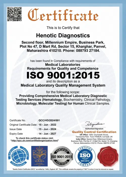
ISO 9001:2015

NABH Standard

PCPNDT Compliant
Official Certification Gallery
In addition, select any certificate below to view the official high-resolution compliance document.
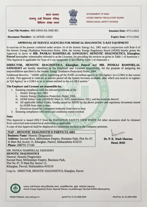



Daily Equipment QA Tolerances & NDRL Dose Tables
Senior Medical Leadership & Consultant Radiologists – Radiology Services
As a result, our radiology services are led by distinguished senior radiologists with extensive subspecialty expertise across neuro-imaging, cross-sectional diagnostics, fetal radiology, and interventional procedures.

Dr. Pratibha Patil, MD (Radiodiagnosis)
Specifically, Dr. In light of this, Pratibha Patil brings over 15 years of clinical mastery in 3.0T MRI, 128-Slice Cardiac CT, Women’s Imaging (BI-RADS 0-6), Fetal Doppler (ISUOG standards), and image-guided non-vascular interventional procedures. Furthermore, she leads an expanded radiologist subspecialty team covering Neuroradiology, Musculoskeletal (MSK) imaging, Fetal/Obstetric Doppler, Body Imaging, and Interventional Radiology to oversee quality assurance, contrast safety, and peer review.
Frequently Asked Questions (FAQ) — Radiology Services
In addition, find comprehensive answers to common patient and referring physician questions regarding diagnostic radiology imaging, appointment preparation, safety protocols, and report interpretation at Henotic Diagnostics.
General Diagnostic Radiology Services FAQs
What are radiology services?
Furthermore, in general, radiology services include diagnostic imaging examinations such as MRI, CT, ultrasound, Doppler, X-ray, mammography, PET-CT and DEXA.
What does a radiologist do?
Specifically, a radiologist is a medical doctor who interprets diagnostic imaging examinations and prepares reports to help referring doctors evaluate and manage medical conditions.
How do I read my radiology diagnostic report?
First, look at the Impression section at the bottom for the radiologist’s primary diagnostic summary. Subsequently, the Findings section contains technical organ descriptions written for your treating physician.
What should parents do if a child is scared of an MRI?
Furthermore, parents can accompany their child into the MRI scanning room. Specifically, Henotic Diagnostics provides 3-minute Fast-MRI HASTE sequences, child-friendly music headphones, and continuous technologist support.
Can I eat right after an IV contrast CT scan?
Yes. Specifically, You may resume your normal diet immediately after your contrast CT scan. In addition, we recommend drinking 1.5 to 2 liters of plain water over the next 24 hours to accelerate natural contrast dye excretion.
Clinical Quality & Reporting FAQs
How do I get a second opinion on an outside radiology scan?
In addition, alternatively, you can bring your outside DICOM CD or upload scan files to our digital PACS portal. Our consultant radiologists perform comprehensive subspecialty over-reads and comparative interpretations.
What is AI-assisted computer-aided detection (CADx) in radiology?
In fact, AI CADx triage algorithms analyze diagnostic scans concurrently with radiologists to automatically highlight critical pathology such as acute intracranial hemorrhage, vessel occlusions, pulmonary emboli, and subtle mammographic microcalcifications.
What are ACR Appropriateness Criteria® guidelines?
Indeed, these are evidence-based guidelines developed by the American College of Radiology to assist referring physicians in choosing the most appropriate diagnostic imaging examination for specific clinical presentations.
What are CAD-RADS, LUNG-RADS, BI-RADS, and PI-RADS lexicons?
In fact, these are standardized, evidence-based reporting frameworks developed by the American College of Radiology to categorize lesion risk, ensure diagnostic clarity, and recommend standardized clinical management.
What is the turnaround time (TAT) for urgent radiology scans?
Consequently, at Henotic Diagnostics, STAT emergency findings are phoned directly to ordering doctors within 15 minutes, urgent inpatient reports are delivered within 1 to 2 hours, and routine screening reports are uploaded to the online PACS portal within 4 to 6 hours.
Specific Radiology Modality FAQs
What are the main types of radiology imaging?
Specifically, in general, the main modalities of radiology imaging are Magnetic Resonance Imaging (MRI), Computed Tomography (CT), Diagnostic Ultrasound & Sonography, Color Doppler Ultrasound, Digital Radiography (X-Ray), Digital Mammography, PET-CT Imaging, Nuclear Medicine, and Bone Mineral Density (DEXA) scans.
Is MRI part of radiology?
Indeed, Magnetic Resonance Imaging (MRI) is an essential non-invasive subspecialty of diagnostic radiology services that utilizes strong magnetic fields and radiofrequency pulses to capture high-resolution cross-sectional soft tissue images without ionizing radiation.
Is ultrasound part of radiology?
Overall, diagnostic ultrasound and Doppler examinations are core components of radiology services that use high-frequency sound waves to visualize internal organs, vascular hemodynamics, and fetal development.
Does CT use radiation?
Yes. Specifically, CT uses X-rays combined with computer processing to create detailed cross-sectional images of the body.
Does MRI use radiation?
No. In fact, MRI does not use ionizing radiation. Moreover, it relies on radio waves and strong magnetic fields.
What is the difference between CT and MRI?
First, CT scans use X-rays for rapid imaging of bony structures, chest pathology, and acute hemorrhages. In contrast, MRI uses magnetic fields to deliver soft tissue contrast resolution for brain, spine, joint, and pelvic evaluations.
Clinical Utility & Exam Selection FAQs
Why is radiology important?
Notably, above all, radiology services provide non-invasive visualization of internal pathology, enabling early disease detection, precise treatment planning, oncological staging, and objective recovery monitoring.
How is an imaging examination selected?
In summary, an imaging test is selected based on clinical symptoms, targeted organ systems, diagnostic sensitivity, safety considerations (such as pregnancy or renal function), and referral guidelines.
Patient Experience, Preparation & Reports FAQs
What happens during a radiology examination?
Additionally, initially, during an imaging exam, certified radiologic technologists position the patient, guide breathing instructions if required, acquire diagnostic scans, and transmit images to radiologists for formal interpretation.
How should I prepare for a radiology examination?
Furthermore, in general, preparation depends on the examination. For example, abdominal ultrasounds and PET-CT scans may require fasting; pelvic ultrasounds require a full bladder; contrast CT/MRI scans require kidney function testing (creatinine); and MRI exams require removing metallic objects.
Who interprets radiology scans?
Finally, diagnostic imaging examinations are interpreted by qualified, licensed radiologists who review the scans and prepare formal diagnostic reports.
Location, Helpline & Operating Hours – Radiology Services
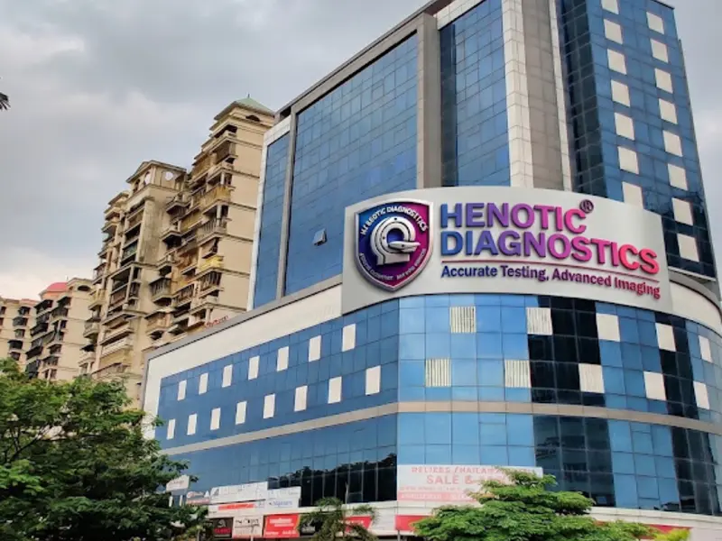
Henotic Diagnostics – Central Kharghar Suite
GIS Geolocation Metadata: Facility coordinates accurately indexed at GIS Geolocation metadata (Latitude: 19.0475 N, Longitude: 73.0699 E) for emergency transport dispatch via Official Google Maps Location Portal.