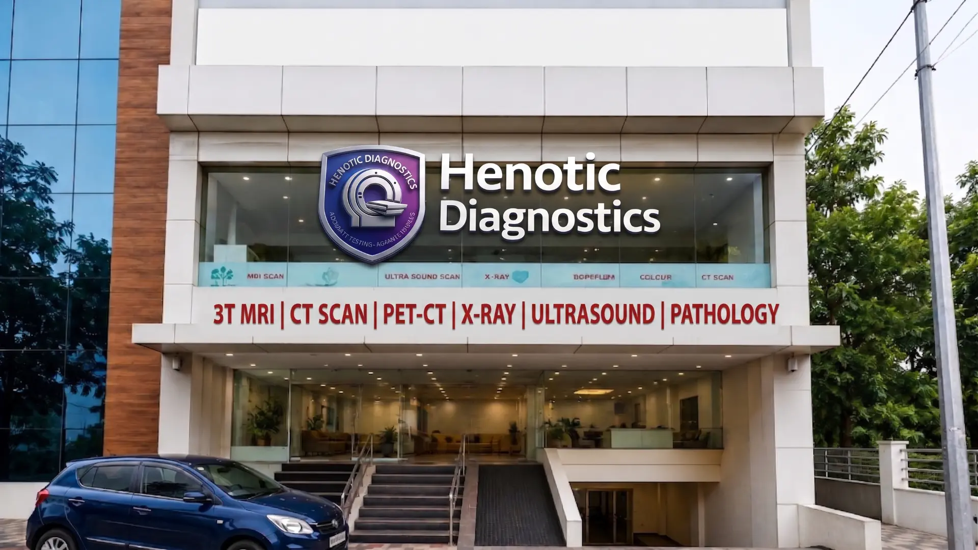
Diagnostic Imaging: Types, Tests, Scans, FFR-CT & Subspecialty Protocols
Fundamentally, diagnostic imaging encompasses non-invasive medical procedures that generate detailed visual representations of internal anatomical structures, tissue micro-architecture, and organ systems. To achieve this, our radiology suite at Henotic Diagnostics features advanced medical technology—including 3.0 Tesla High-Field MRI, 128-Slice Low-Dose CT, diagnostic 3D/4D Ultrasound, Amyloid & 68Ga-PSMA PET-CT, 3D Digital Breast Tomosynthesis, and computational FFR-CT cardiac imaging. Furthermore, all diagnostic examinations adhere strictly to ALARA (As Low As Reasonably Achievable) radiation safety protocols as well as AERB DRL benchmarks, while every scan is interpreted directly by experienced consultant radiologists.
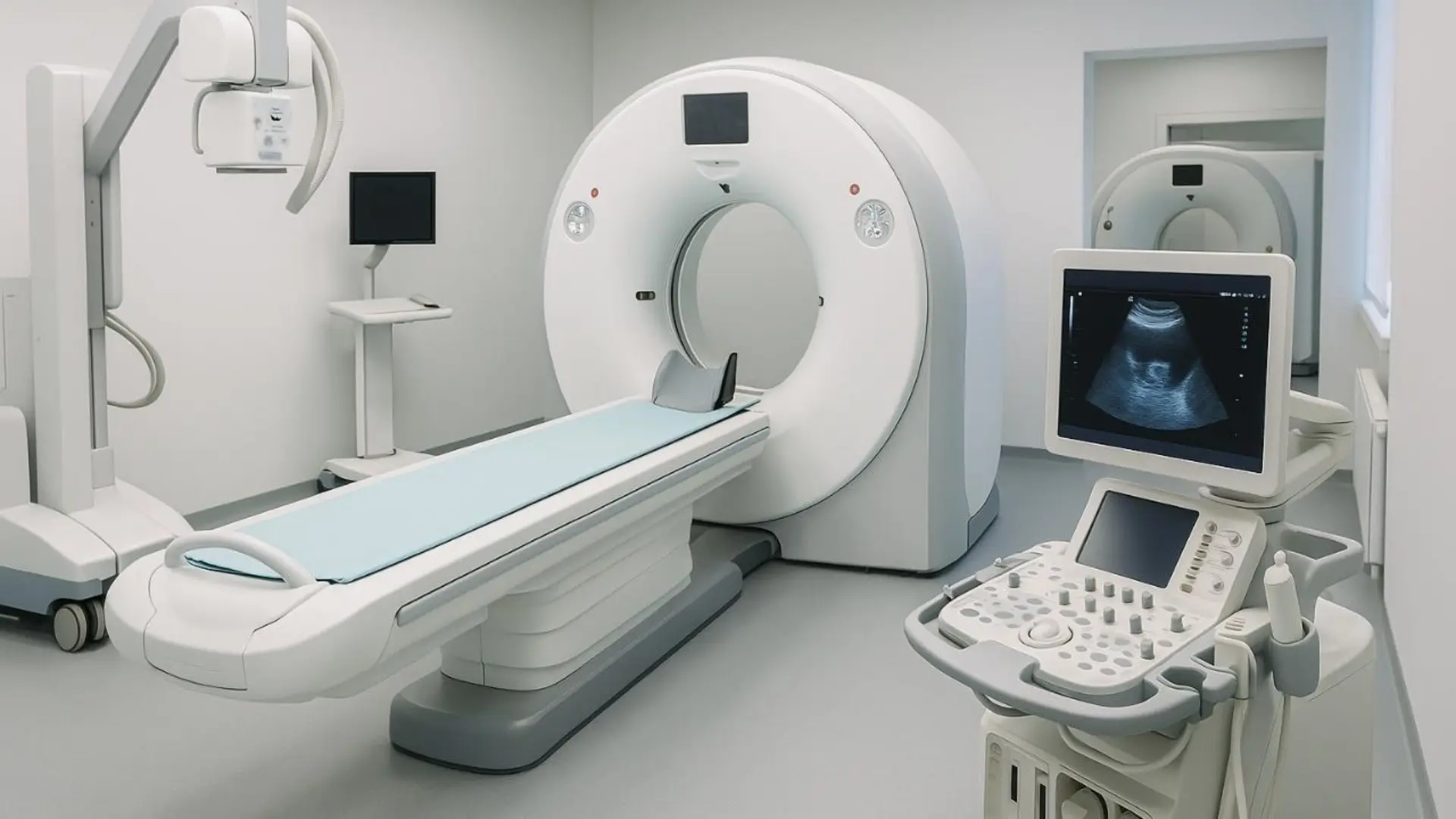
Diagnostic Imaging Modalities & Technical Capabilities
Remarkably, our center integrates high-field magnetic resonance, ultra-fast volumetric multi-detector CT, high-frequency sound wave sonography, and functional metabolic PET-CT within a unified diagnostic facility. Consequently, patients receive rapid diagnostic evaluations under one roof.
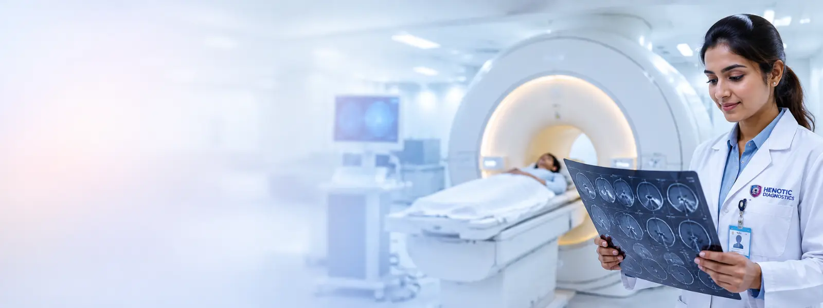
High-Field 3T MRI Diagnostic Imaging
Specifically, high-resolution soft-tissue contrast is acquired using nuclear magnetic resonance without ionizing radiation. In addition, advanced sequences include Neuromelanin-Sensitive (NM-MRI) Parkinson’s imaging, BOLD Functional fMRI motor/language mapping, GI-RADS deep endometriosis MRI, 3D HR-VWI vessel wall imaging, and mpMRI prostate staging.
Explore 3T MRI Scan Services →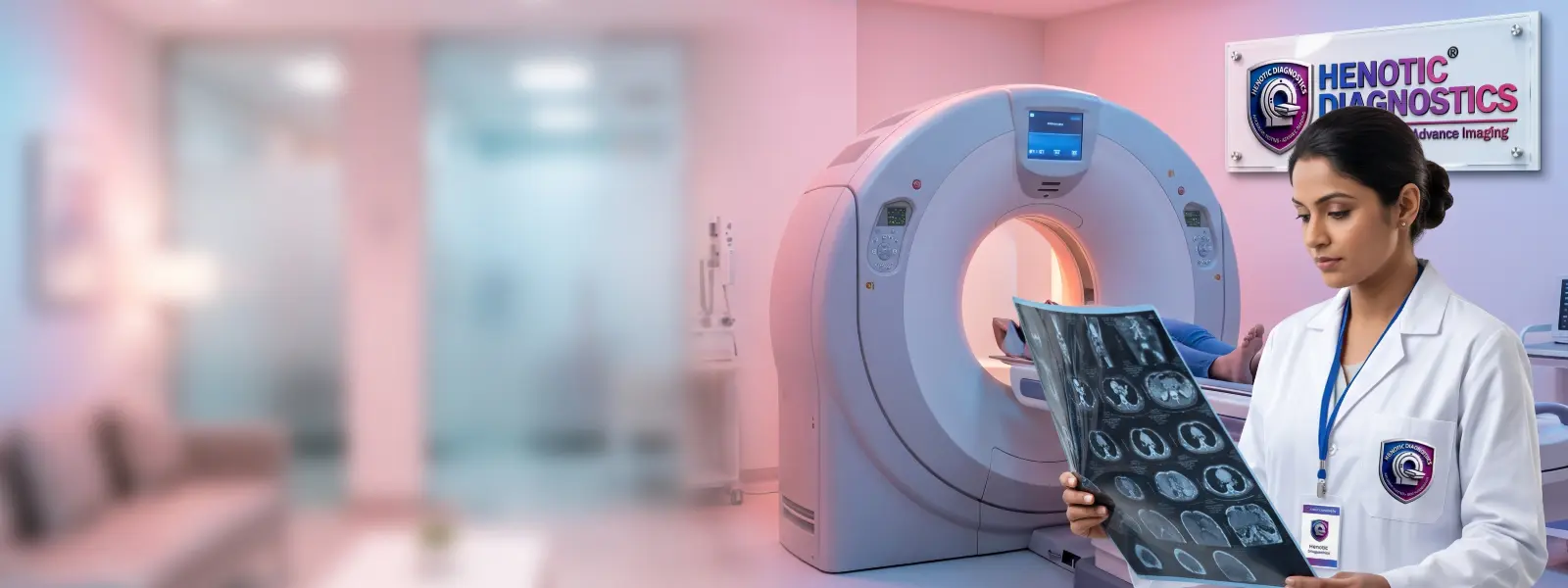
128-Slice Volumetric CT Diagnostic Imaging
Furthermore, ultra-fast sub-millimeter cross-sectional imaging is powered by computational FFR-CT flow dynamics. As a result, specialized features include Dynamic CT Myocardial Perfusion (CTMP), Spectral Dual-Energy CT (Z-eff stone composition), emergency Triple-Rule-Out (TRO) chest pain CT, and CT Angiography.
Explore 128-Slice CT Services →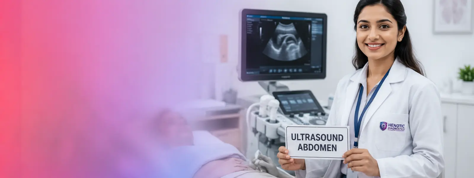
Diagnostic Ultrasound & Sonography
Additionally, real-time Tissue Harmonic Imaging (THI) operates using non-ionizing sound waves. For instance, diagnostic protocols encompass Neonatal Transfontanellar Neuro-Sonography, ISUOG fetal neurosonography, Dynamic MSK Stress Sonography, 2D Shear Wave Elastography (SWE in kPa), and USG-guided interventional procedures.
Explore Ultrasound Scan Services →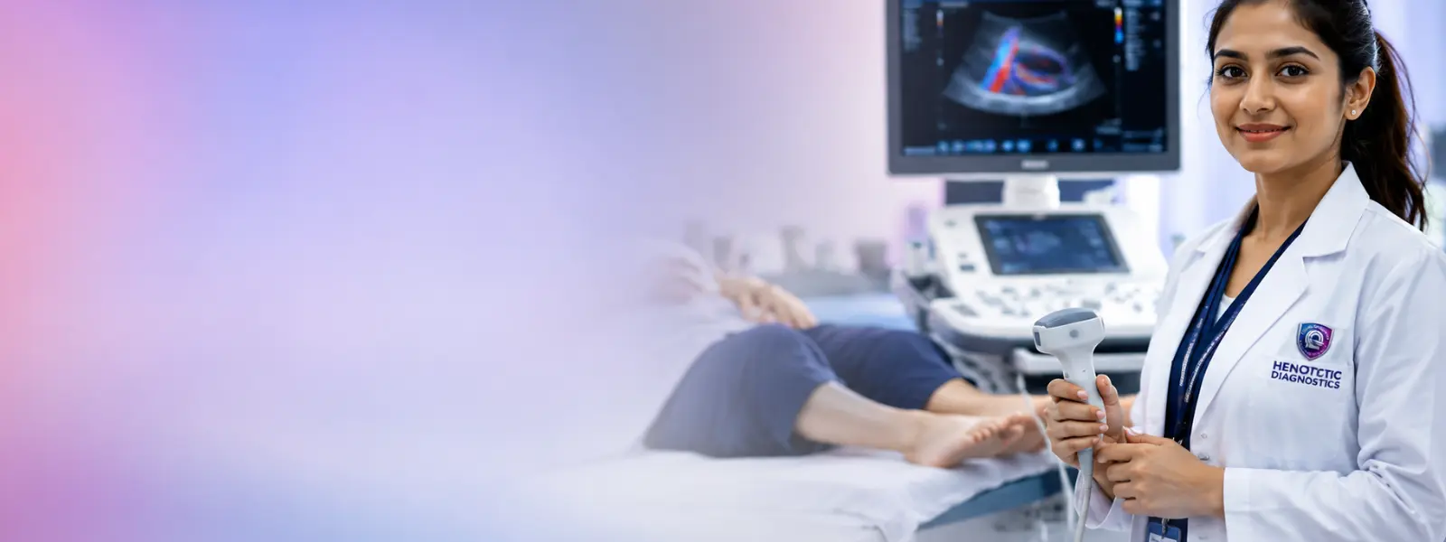
Color Doppler Diagnostic Imaging
Meanwhile, specialized hemodynamic vascular sonography analyzes Peak Systolic Velocity (PSV) and Deep Vein Thrombosis (DVT). Consequently, clinicians receive accurate assessments for V-RADS endoleak monitoring and carotid arterial stenosis.
Explore Color Doppler Services →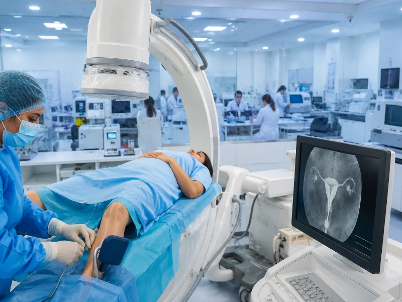
Digital Radiography (X-Ray Diagnostic Imaging)
Moreover, ultra-low-dose flat-panel digital radiography supports Hysterosalpingography (HSG) testing by delivering rapid anatomical images. As a result, physicians quickly assess fallopian tube patency, uterine abnormalities, pelvic structures, reproductive anatomy, and related gynecological conditions.
Explore Digital Radiography Services →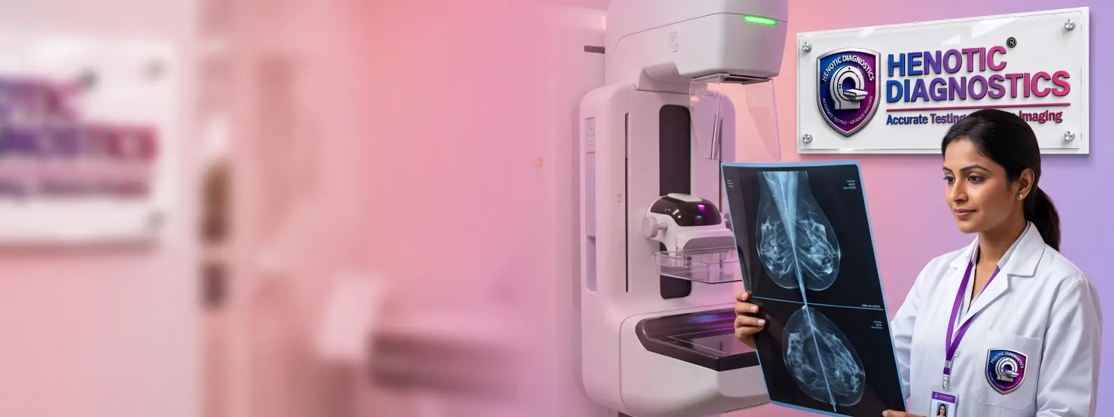
Diagnostic Breast Imaging & 3D Mammography
In particular, Mammography, Digital Mammography, 3d Mammography, Sonomammography and Digital Breast Tomosynthesis utilizes Modulation Transfer Function spatial frequency optics. Consequently, this technology enables early microcalcification detection and precise architectural distortion analysis.
Explore Mammography Services →
Theranostic PET-CT Diagnostic Imaging
Furthermore, molecular radiotracer imaging features Amyloid 18F-Flutemetamol for early Alzheimer’s plaque mapping. Additionally, we provide 68Ga-PSMA for prostate cancer and post-therapy 177Lu radieligand dosimetry (Gy).
Explore PET-CT Scan Services →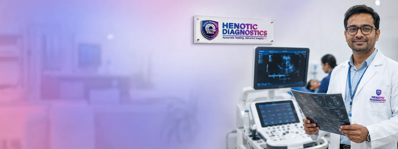
Cardiac Diagnostic Imaging Services
Likewise, non-invasive cardiac evaluations combine 2D Echocardiography, 4D Flow CMR Wall Shear Stress (WSS), dynamic CTMP perfusion, and 128-slice FFR-CT Coronary Angiography for non-invasive pressure drop calculations.
Explore Cardiac Imaging Services →Need Assistance Selecting the Right Diagnostic Scan?
Specifically, our radiology team actively assists referring clinicians and patients. For example, we systematically verify scan protocols, contrast requirements, and pre-test preparation guidelines.
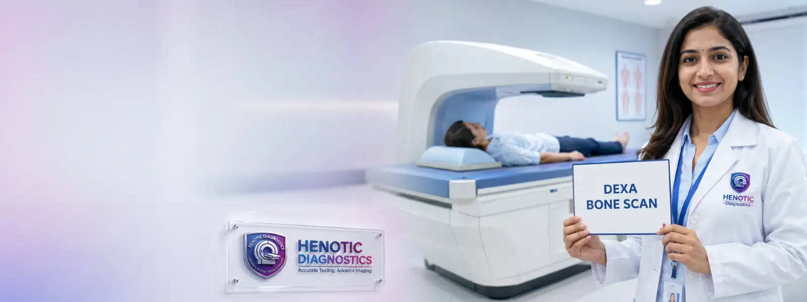
DEXA Bone Densitometry Diagnostic Imaging
Additionally, Dual-energy X-ray absorptiometry measures bone mineral density (BMD) at lumbar spine and hip regions. Therefore, clinicians seamlessly integrate FRAX 10-year fracture risk estimations.
Explore DEXA Bone Density Scans →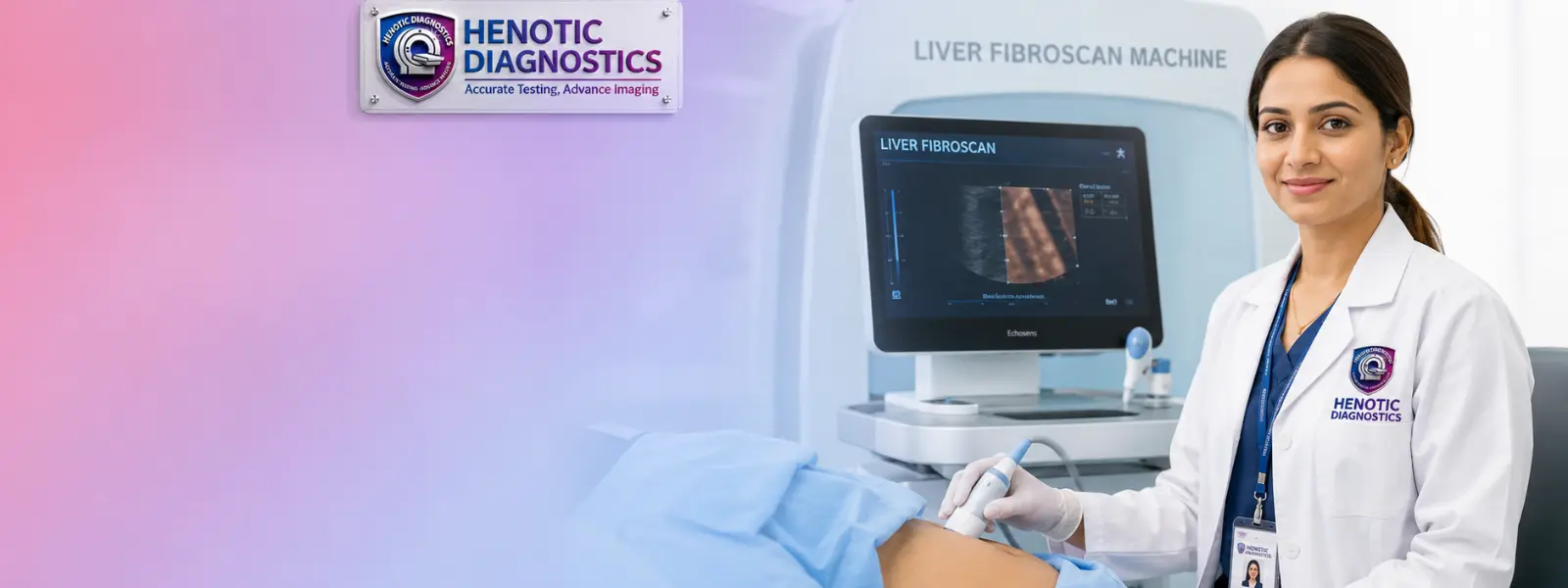
FibroScan Diagnostic Imaging & Liver Elastography
Simultaneously, non-invasive transient elastography measures liver stiffness (in kilopascals, kPa) alongside Controlled Attenuation Parameter (CAP in dB/m). Consequently, this provides comprehensive Cirrhosis Screening, Chronic Liver Disease Screening, hepatic steatosis and fibrosis staging.
Explore FibroScan Services →What Is Diagnostic Imaging? Physical & Physiological Basis
Definition & Scope of Diagnostic Imaging
Fundamentally, diagnostic imaging encompasses non-invasive medical modalities that record spatial, anatomical, and functional energy maps of human tissue. As a result, these procedures enable physicians to evaluate structural integrity, detect silent pathologies, and monitor medical treatments without surgical intervention.
Medical Physics & Physics Principles in Diagnostic Imaging
Specifically, each diagnostic imaging modality operates on distinct physical principles to differentiate pathological tissue from surrounding normal architecture:
- Magnetic Resonance Imaging (MRI Physics & K-Space): First, high-strength static magnetic fields (3.0 Tesla) align hydrogen nuclear dipoles (protons). Subsequently, radiofrequency (RF) pulses excite protons, and as they relax back to baseline, spatial frequency data is stored in K-Space. Ultimately, Fast Fourier Transformation (FFT) converts K-space matrix raw data into anatomical slice images, while T1 and T2 relaxation times dictate contrast.
- Computed Tomography (CT Physics & ASIR-V): Second, a rotating X-ray tube emits fan-beam radiation measured across 128 detector rows. Consequently, tissue attenuation is converted into Hounsfield Units (HU). In addition, Model-Based Iterative Reconstruction (ASIR-V) algorithms reconstruct volumetric images while reducing radiation exposure by up to 70%.
- Diagnostic Ultrasound & Doppler Physics: Third, transducers convert electrical impulses into acoustic waves (2–15 MHz). Meanwhile, reflections at acoustic impedance boundaries generate grayscale images. Furthermore, the Doppler Effect measures frequency shifts in moving erythrocytes to calculate hemodynamics via Doppler Frequency Shift: Δf = (2f₀v cosθ) / c.
- Positron Emission Tomography (PET-CT): Finally, 18F-FDG radiotracers emit positrons that annihilate with surrounding electrons, releasing paired 511 keV gamma photons. Consequently, coincidence detectors capture these emissions to measure glucose metabolic hotspots (SUV max).
Diagnostic Imaging vs. Laboratory Pathology
Whereas clinical laboratory pathology measures biochemical biomarkers, serum enzymes, and cellular counts, diagnostic imaging provides three-dimensional spatial localization, morphological staging, and vascular assessment. Therefore, combining laboratory metrics with imaging findings forms the foundation of modern evidence-based medicine.
FFR-CT Computational Fluid Dynamics & CMR 4D Flow WSS
Fractional Flow Reserve by CT (FFR-CT)
Specifically, by applying 3D Computational Fluid Dynamics (CFD) and aortic root boundary pressure conditions to 128-slice CT Coronary Angiograms, FFR-CT non-invasively calculates functional pressure drops (FFR-CT ≤ 0.80 indicating ischemia). Consequently, this eliminates unnecessary invasive cardiac catheterizations for stable coronary artery disease.
4D Flow CMR Wall Shear Stress (WSS) & TKE
Similarly, phase-contrast 4D Flow Cardiac MRI maps 3D spatial velocity vectors over time. As a result, it accurately quantifies Wall Shear Stress (WSS) and Turbulent Kinetic Energy (TKE) in thoracic aortic aneurysms, bicuspid aortic valves, and portal vein flow dynamics.
Neuromelanin-Sensitive 3T MRI & BOLD Functional fMRI
Neuromelanin-Sensitive 3T MRI (NM-MRI)
Specifically, a high-resolution T1-weighted 3T sequence evaluates substantia nigra pars compacta and locus coeruleus depigmentation. Consequently, this enables early differential diagnosis of Parkinson’s Disease versus Progressive Supranuclear Palsy (PSP) prior to structural basal ganglia atrophy.
BOLD Functional MRI (fMRI) Eloquent Cortex Mapping
In contrast, Blood Oxygen Level-Dependent (BOLD) task-based fMRI maps primary motor cortex, sensory strips, and Broca’s/Wernicke’s speech centers relative to intracranial brain tumors. Therefore, neurosurgeons effectively utilize these maps to guide operative trajectories.
Pediatric Transfontanellar Sonography & Fetal Neurosonography
- Neonatal Transfontanellar Neuro-Sonography: Specifically, high-frequency ultrasound through the anterior fontanelle assesses neonatal Germinal Matrix Hemorrhages (Grades I–IV), periventricular leukomalacia (PVL), and hypoxic-ischemic encephalopathy (HIE) without sedation.
- Fetal Neurosonography (ISUOG Guidelines): In addition, multi-planar 3D/4D sonography systematically evaluates fetal corpus callosum dysgenesis, posterior fossa cisterns, and cerebellar vermis integrity.
Amyloid PET-CT & Radieligand Theranostic Dosimetry
18F-Flutemetamol Amyloid PET
Specifically measures in vivo quantitative cortical beta-amyloid plaque mapping for early Alzheimer’s disease diagnosis.
177Lu Theranostic Dosimetry
Subsequently calculates post-therapy SPECT-CT/PET-CT absorbed dose (Gy) for salivary glands, kidneys, and bone marrow.
GI-RADS Endometriosis MRI
Additionally, it accurately evaluates Deep Infiltrating Endometriosis (DIE) staging of uterosacral ligaments and rectosigmoid junction on 3T MRI.
V-RADS Vascular Reporting
Furthermore, it systematically provides standardized reporting for EVAR endoleaks (Types I–V) and peripheral arterial disease staging.
Diagnostic Reference Levels (DRLs) & Radiation Registry
In accordance with AERB guidelines, Henotic Diagnostics systematically operates automated institutional radiation dose registries:
How Is the Right Diagnostic Imaging Test Selected? (ACR Guidelines)
First and foremost, diagnostic scan selection at Henotic Diagnostics strictly follows established ACR (American College of Radiology) Appropriateness Criteria and ICRI guidelines. Specifically, referring physicians select tests based on precise clinical decision variables:
- Anatomical Region & Target Tissue: Primarily, neural tissue, ligaments, and menisci mandate high soft-tissue contrast MRI. In contrast, cortical bone fractures and pulmonary alveoli mandate high-spatial CT or digital X-ray.
- Emergency STAT Triage: Meanwhile, acute trauma, suspected stroke, pulmonary embolism, or acute aortic dissection demand ultra-fast multidetector CT evaluation with sub-hour report turnaround.
- Renal Function Screening (eGFR Thresholds): In addition, contrast-enhanced CT or MRI requires serum creatinine screening. Specifically, an eGFR ≥ 60 mL/min/1.73m² is standard for IV contrast administration, whereas patients with eGFR < 30 mL/min/1.73m² undergo alternative non-contrast protocols.
- Pregnancy Status & Radiation Avoidance: Finally, pregnant patients receive non-ionizing Ultrasound or non-contrast MRI (after first-trimester review) to eliminate fetal ionizing radiation exposure.
Patient Comfort & Safety During Diagnostic Imaging Procedures
First and foremost, we prioritize patient safety, anxiety reduction, and strict pre-scan screening across all imaging suites:
MRI Diagnostic Imaging Implant Safety
Due to the high-strength 3.0 Tesla magnetic field, all implants are systematically verified against safety standards prior to suite entry:
- Certified MR Safe: First, non-conducting, non-metallic items (such as plastic or silicon) are completely safe in all magnetic environments.
- Verified MR Conditional: Second, implants requiring specific field strength verification (such as specific cardiac pacemakers, artificial heart valves, or joint replacements) must be checked. Consequently, official implant documentation must be provided prior to scanning.
- Strictly MR Unsafe: Third, items hazardous in magnetic fields (including older ferromagnetic aneurysm clips, metallic ocular fragments, or non-certified cochlear implants) are strictly prohibited.
Claustrophobia Management in Diagnostic Imaging Scans
Additionally, patients experiencing claustrophobia or scan-related anxiety receive supportive care. For instance, we offer wide-bore scanner gantries, ambient LED lighting, continuous noise-reduction headphones, two-way intercom communication, and squeezable emergency call bulbs. Furthermore, accompaniment by a family member is supported following safety screening.
Diagnostic Imaging Preparation Guidelines by Modality
| Modality | Fasting (NPO) Window | Hydration & Bladder Requirements | Clothing & Metallic Screening |
|---|---|---|---|
| MRI (Non-Contrast) | None required | Normal hydration | First, remove all jewelry, metal zippers, wigs, and metallic cosmetics before changing into a medical gown. |
| Contrast CT / MRI | 4 – 6 hours solid food fasting | Hydrate with 1 Liter water before test | Specifically, a recent serum creatinine report (< 30 days) is required prior to contrast injection. |
| Abdominal Ultrasound | 6 hours strict fasting | Sip water; avoid carbonated drinks | As a result, fasting reduces bowel gas interference for gallbladder & liver visualization. |
| Pelvic / Obstetric USG | None required | 1 Liter water 1 hour prior (Full Bladder) | Consequently, a full urinary bladder creates an acoustic window to view pelvic organs. |
Diagnostic Imaging Protocols During Pregnancy
Protecting maternal and fetal health fundamentally requires adherence to radiation safety guidelines established by ACOG (American College of Obstetricians and Gynecologists) and RCR:
- Diagnostic Ultrasound (Primary Modality): First, high-frequency sound waves carry zero ionizing radiation risk. Consequently, acoustic output parameters—such as Thermal Index (TI) and Mechanical Index (MI)—are kept well below statutory limits.
- MRI During Pregnancy: Second, high-field non-contrast MRI is considered safe during the second and third trimesters when diagnostic information cannot be obtained via ultrasound. However, gadolinium contrast is strictly avoided during pregnancy.
- X-Ray and CT Precautions: Third, examinations involving ionizing radiation to the abdomen or pelvis are avoided during pregnancy unless life-threatening clinical emergencies arise. Accordingly, abdominal lead shielding is mandatory for essential non-abdominal X-rays.
Contrast Media Safety in Diagnostic Imaging Scans
Specifically, intravenous contrast media significantly enhances diagnostic sensitivity by highlighting vascular anatomy and lesion margins:
- Non-Ionic Low-Osmolar Iodinated Contrast (CT Scans): Modern non-ionic agents significantly reduce physiological osmolality risks, thereby lowering hypersensitivity reaction rates.
- Gadolinium-Based Contrast Agents (GBCA – MRI Scans): In addition, macrocyclic gadolinium agents are selected to minimize tissue retention risks.
- Contrast-Induced Nephropathy (CIN) Prevention: Furthermore, renal function (eGFR) is evaluated prior to contrast injection. Consequently, high-risk patients receive pre-scan and post-scan IV hydration protocols.
- Emergency Readiness: Finally, our facility maintains emergency resuscitation equipment, oxygen support, and anti-histamine/epinephrine management protocols under direct physician supervision.
Understanding Your Diagnostic Imaging Report & RADS Systems
Specifically, radiology reports at Henotic Diagnostics are systematically structured according to international standardized reporting frameworks:
Standardized RADS Systems in Diagnostic Imaging
BI-RADS (Breast)
Specifically, Breast Imaging Reporting and Data System (Categories 0–6) standardizes mammography findings from benign to biopsy-indicated guided.
PI-RADS (Prostate)
Additionally, Prostate Imaging Reporting and Data System (Scores 1–5) assesses clinically significant prostate cancer risk on multiparametric MRI.
TI-RADS (Thyroid)
Furthermore, Thyroid Imaging Reporting and Data System categorizes ultrasound nodule features to guide fine-needle aspiration (FNAC).
LI-RADS (Liver)
Similarly, Liver Imaging Reporting and Data System categorizes focal hepatic lesions in high-risk liver cirrhosis patients.
Diagnostic Imaging Technical Glossary
| Radiological Term | Modality | Clinical Meaning |
|---|---|---|
| Hyperintense / Hypointense | MRI | Specifically, bright (hyper) or dark (hypo) signal intensity appears on specific sequences (T1, T2, FLAIR). |
| Hypoattenuating / Hyperattenuating | CT Scan | In contrast, tissue absorbs fewer X-rays (darker/fluid) or more X-rays (brighter/calcified) measured in Hounsfield Units. |
| Diffusion Restriction (High DWI / Low ADC) | MRI | Meanwhile, restricted water molecule movement indicates acute ischemic stroke, dense cellular tumors, or abscesses. |
| Radiopaque / Radiolucent | Digital X-Ray | Consequently, structures block X-rays (white/bones) versus structures allowing passage (dark/air-filled lungs). |
Limitations & Artifacts in Diagnostic Imaging Studies
While modern imaging modalities offer diagnostic clarity, radiologists carefully account for physical limitations and imaging artifacts during interpretation:
- Motion Artifacts: Involuntary patient movement or cardiac motion can blur image fine detail, thereby requiring fast pulse sequences or breath-holding protocols.
- Beam Hardening & Metal Artifacts: In addition, dense metal implants cause streak artifacts on CT and magnetic susceptibility signal loss on MRI. Consequently, advanced metal artifact reduction (MAR) software is used to minimize distortion.
- Clinical-Pathological Correlation Required: Specifically, imaging findings reflect structural and metabolic alterations. Ultimately, final diagnostic conclusions require correlation with clinical history, physical examination, and histopathological biopsy when indicated.
Digital Diagnostic Imaging PACS & Cloud Report Access
First and foremost, diagnostic image data acquired at Henotic Diagnostics is systematically processed through enterprise PACS (Picture Archiving and Communication System). Consequently, high-resolution DICOM image datasets and verified digital reports are accessible online for patients and treating specialists, thereby supporting rapid clinical decision-making and second-opinion reviews.
Diagnostic Imaging Protocols by Body Region & Medical Specialty
Brain & Neuro Diagnostic Imaging
Specifically, 3T Neuro-MRI (DWI/ADC, FLAIR, SWI) evaluates stroke, demyelination, and intracranial mass lesions, whereas CT Head assesses acute intracranial hemorrhage.
Spine & Spinal Cord Diagnostic Imaging
Additionally, Cervical and Lumbar Spine MRI evaluate disc herniation and spinal canal stenosis, while CT Spine provides complex fracture mapping.
Chest & Pulmonary Diagnostic Imaging
Furthermore, HRCT Chest (1mm thin cuts) evaluates interstitial lung disease (ILD), while digital chest radiography assesses parenchymal consolidation.
Gastrointestinal Diagnostic Imaging
Meanwhile, Abdominal Ultrasound evaluates cholelithiasis, whereas contrast Triple-Phase CT and non-invasive 3D MRCP provide detailed hepatic and pancreaticobiliary evaluation.
Pelvic & Prostate Diagnostic Imaging
In addition, Pelvic Ultrasound and dedicated PI-RADS Multiparametric MRI evaluate uterine fibroids, ovarian cysts, and prostate disease.
Diagnostic Breast Imaging & Mammography
Likewise, 3D Tomosynthesis Mammography (BI-RADS) combines with high-frequency Breast Ultrasound for comprehensive dense tissue evaluation.
Diagnostic Imaging Across Medical Specialties & Clinical Workflows
Our radiologist reporting strictly aligns with subspecialty requirements, thereby supporting multidisciplinary clinical workflows:
Oncology Diagnostic Imaging & RECIST 1.1
Specifically, PET-CT and contrast-enhanced CT scans are interpreted using standardized RECIST 1.1 (Response Evaluation Criteria in Solid Tumors) to measure target lesion response following chemotherapy or radiotherapy.
Orthopedic & Musculoskeletal Diagnostic Imaging
In addition, high-field 3T MRI sequences evaluate joint biomechanics, thereby grading ligament and tendon injuries from Grade I (micro-tears) to Grade III (complete tear with retraction).
Neurological Diagnostic Imaging & Stroke Protocols
Furthermore, neuro-imaging integrates DWI/ADC stroke protocols and CT Angiography to evaluate penumbra tissue viability and large vessel occlusions (LVO) for hyperacute intervention.
Why Choose Diagnostic Imaging at Henotic Diagnostics
Sub-Millimeter Advanced Hardware
Specifically, high-field 3T MRI, 128-slice volumetric low-dose CT, 3D Tomosynthesis, and 18F-FDG PET-CT are housed within a single facility.
Board-Certified Radiologist Leadership
Additionally, diagnostic reports are directly authored and reviewed by consultant radiologists experienced in cross-sectional imaging.
Statutory Radiation & Quality Compliance
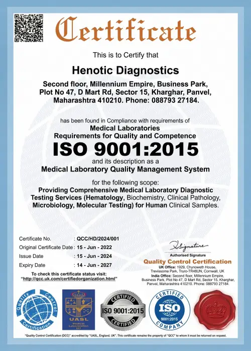
Furthermore, we maintain full statutory compliance with AERB radiation safety norms, PCPNDT regulations, NABL ISO 15189, and ISO 9001:2015 quality standards.
STAT Emergency Reporting & PACS Portal
As a result, we provide sub-hour STAT reporting turnarounds for emergency cases alongside cloud DICOM image access for treating specialists.
Diagnostic Imaging Reporting Radiologist & Medical Leadership
Specifically, diagnostic imaging interpretation at Henotic Diagnostics Radiology Center is led by our consultant radiologist.

Dr. Pratibha Patil
Consulting Radiologist | MD / DNB Radiology
Specifically, Dr. Patil brings over 15 years of subspecialty experience in diagnostic cross-sectional imaging, 3T MRI interpretation, multi-detector CT angiography, and diagnostic sonography.
In this role, Dr. Pratibha Patil oversees the radiology diagnostic reporting workflow at Henotic Diagnostics.
Furthermore, her clinical focus encompasses neuro-radiology MRI, CT angiography, pelvic sonography, and multi-modality cancer imaging.
Consequently, working alongside referring physicians, Dr. Patil ensures that every imaging study provides clear diagnostic findings to assist patient care.
Diagnostic Imaging Accreditations & Regulatory Compliance
Specifically, diagnostic imaging quality and safety standards at Henotic Diagnostics align with national regulatory authorities and quality frameworks.
Diagnostic Imaging Accreditations & Authorities
AERB Compliant
Radiation Safety & Diagnostic Equipment Quality Assurance

NABL ISO 15189
Laboratory Quality & Diagnostic Competency Accreditation

ISO 9001:2015
International Quality Management System Certified

ISUOG Member
International Society of Ultrasound in Obstetrics & Gynecology

NABH Compliant
Healthcare Facility Quality & Patient Safety Protocols

PCPNDT Registered
Full Compliance with Pre-Conception & Pre-Natal Diagnostic Techniques Act
Official Diagnostic Imaging Certifications



Patient Experiences with Diagnostic Imaging Services
Specifically, read patient experiences regarding communication, scheduling, and diagnostic reporting at Henotic Diagnostics.
Verified Patient Experiences with Diagnostic Imaging Scans
“Recently, I underwent an MRI spine scan at Henotic Diagnostics. Furthermore, the MRI room was clean, the staff explained the procedure clearly, and the digital report was delivered to my doctor promptly.”
— Rajesh M., Kharghar
“When my doctor requested an abdominal ultrasound and Doppler study, the sonographer was gentle, efficient, and ensured my comfort throughout the examination.”
— Sunita S., Navi Mumbai
“Overall, the CT scan procedure was fast and well-coordinated. Consequently, receiving digital report access on my phone saved me an extra trip to collect physical films.”
— Amit K., Panvel
“My experience with the diagnostic imaging team was smooth and comfortable. The staff were professional, explained the scan process clearly, and provided the report within the expected timeframe.”
— Neha P., Kamothe