MRI Ankle Scan: Advanced Soft Tissue Diagnostic Imaging
Experience the absolute highest standard of clinical clarity. Our cutting-edge, high-resolution joint neuroimaging technology ensures precise evaluation of chronic foot pain, complex ligament tears, Achilles tendon disorders, and hidden structural abnormalities. Consequently, you can confidently obtain rapid, reliable results to fast-track your recovery plan.
Trust Signals: NABL Quality Guidelines | Advanced 3.0T Technology | Expert Radiologists
WhatsApp Appointment | Call 088793 27184
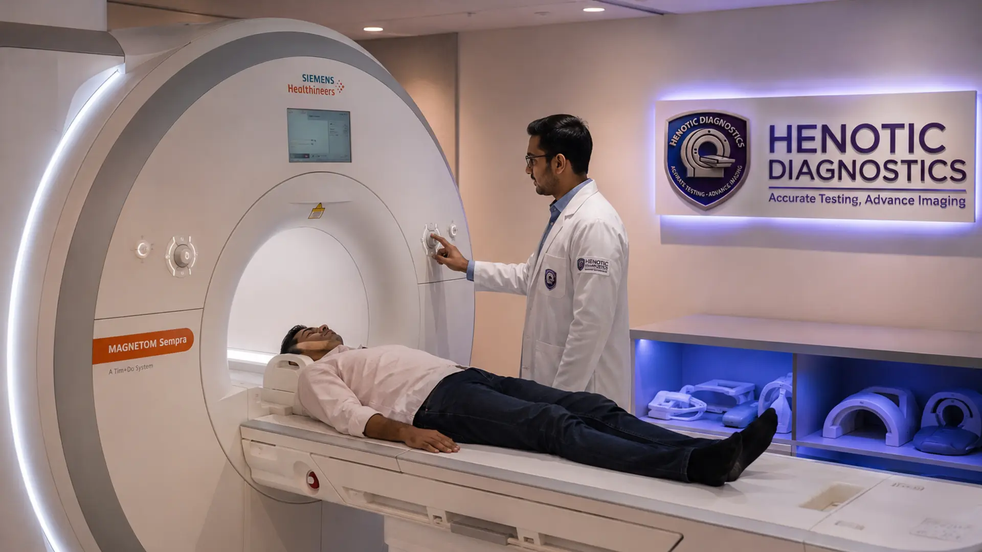
What is an Ankle MRI Scan?
An MRI Ankle scan is a non-invasive diagnostic technique that utilizes a remarkably powerful magnetic field and radiofrequency pulses. Consequently, this state-of-the-art system generates highly detailed multiplanar images across axial, coronal, and sagittal views without exposing you to harmful radiation.
[mri_brain_swiper]Furthermore, physicians favor this imaging protocol whenever they suspect internal derangement. Unlike routine standard X-rays, magnetic resonance imaging provides exceptional soft tissue visualization. Meanwhile, it captures subtle variations within deep structures, which significantly enhances total diagnostic accuracy for medical specialists worldwide.
Why MRI is the Best Imaging Test for the Ankle
To understand why orthopedic surgeons prefer MRI, we must compare it against other standard imaging modalities.
| Feature | MRI Scan | CT Scan | Ultrasound | Digital X-Ray |
|---|---|---|---|---|
| Soft Tissue Detail | Excellent | Poor | Moderate | None |
| Bone Marrow Edema | Gold Standard | Invisible | Invisible | Invisible |
| Radiation Risk | Zero | High | Zero | Low |
| Cartilage Visibility | Highly Detailed | Limited | Obstructed | Joint Space Only |
Anatomy of the Ankle Visualized via MRI
Our expert radiologists use this technology to map every intricate component of your lower joint.
Bony Structures
- Distal Tibia and Fibula
- Talus and Calcaneus (Heel bone)
- Navicular, Cuboid, and Cuneiforms
Ligament Complexes
- Anterior Talofibular Ligament (ATFL)
- Calcaneofibular Ligament (CFL)
- Posterior Talofibular Ligament (PTFL)
- Deltoid and Syndesmotic Ligaments
Tendons and Soft Tissues
- Achilles Tendon
- Tibialis Posterior and Anterior Tendons
- Peroneal and Flexor Tendons
- Plantar Fascia and Articular Cartilage
What Does an MRI Ankle Detect?
This comprehensive scan isolates exact structural failures. Therefore, it effectively identifies the following pathologies:
- Ligament Injuries: ATFL tears, CFL tears, and deep deltoid sprains.
- Tendon Disorders: Achilles tendon rupture, tendinosis, and peroneal tendon tears.
- Bone Disorders: Stress fractures, bone bruises (contusions), and osteochondral lesions of the talar dome.
- Joint & Inflammatory Conditions: Synovitis, joint effusions, osteoarthritis, and plantar fasciitis.
- Masses & Infections: Ganglion cysts, osteomyelitis (bone infection), abscesses, and soft tissue tumors.
- Nerve Entrapment: Tarsal tunnel syndrome and Morton’s neuroma.
Symptoms That May Require an Ankle MRI
You should consult your doctor and consider advanced imaging if you experience any of these persistent symptoms:
- Severe, unrelenting heel or foot pain
- Chronic swelling that fails to subside with ice and rest
- A distinct popping or snapping sound during a twisting injury
- Joint instability or a sensation that the ankle is “giving way”
- Stiffness, locking, or clicking when walking
- Numbness, tingling, or a burning sensation indicating nerve damage
MRI Protocol: With Contrast vs. Without Contrast
Depending on your specific clinical history, your referring physician will request one of two primary approaches.
| Protocol Type | When It Is Used | Key Advantages |
|---|---|---|
| Without Contrast (Standard) | Sports injuries, sprains, tears, and fractures. | Fast, non-invasive, and highly accurate for over 90% of routine orthopedic injuries. |
| With Contrast (Gadolinium) | Tumors, deep infections, and post-surgical evaluations. | Highlights abnormal hyper-vascular tissues, exposing hidden infectious borders and masses. |
Preparation Before Your MRI
Following these steps ensures a safe and comfortable imaging experience.
- Screening: Inform our staff about any pacemakers, cochlear implants, surgical screws, or vascular stents.
- Clothing: Wear loose, comfortable, metal-free clothing. We will provide a medical gown if necessary.
- Jewelry: Remove all earrings, watches, piercings, and metallic accessories before entering the magnetic zone.
- Fasting: Fasting is generally not required unless you are scheduled for a contrast-enhanced scan.
- Documentation: Bring previous X-rays, ultrasound reports, and your doctor’s referral notes.
The MRI Procedure: What to Expect
The examination is entirely painless and straightforward.
- Our technologist will position you comfortably on the motorized scanner table.
- We will place a specialized receiver coil around your ankle to capture localized, high-resolution signals.
- The table slides into the MRI tube. You must remain completely still during the process to avoid motion artifacts.
- The machine will make loud tapping and thumping noises. We provide earplugs or headphones for your comfort.
- The entire scanning process typically lasts between 20 to 35 minutes.
Understanding Your MRI Findings
Once the scan completes, our consultant radiologist evaluates the sequences. Here is what common report terms mean:
- Bone Marrow Edema: Indicates fluid buildup inside the bone, usually pointing to a micro-fracture or acute stress response.
- Grade I Sprain: Minor stretching of the ligament with no gross tearing.
- Grade II Sprain: A partial tear of the ligament causing moderate joint instability.
- Grade III Sprain: A complete rupture of the ligament requiring intensive therapy or surgical intervention.
- Joint Effusion: Excess synovial fluid inside the joint capsule, signaling active inflammation or trauma.
Frequently Asked Questions
Does an ankle MRI use radiation?
No. MRI utilizes powerful magnetic fields and radio waves, meaning it is 100% radiation-free and inherently safe.
How long does an ankle MRI take?
A standard ankle scan requires approximately 20 to 35 minutes. However, if your doctor requests a contrast injection, it may take up to 45 minutes.
Can an MRI detect nerve damage in the foot?
Yes. High-resolution multiplanar imaging sequences clearly visualize nerve entrapment zones, deep neuromas, and conditions like tarsal tunnel syndrome.
Can pregnant women undergo an ankle MRI?
While MRI involves no radiation, we typically avoid scanning during the first trimester as a precaution. Always consult your obstetrician prior to booking.
When will I receive my MRI report?
Our expert radiologists process and deliver comprehensive, cross-verified structural reports within 24 hours of scan completion.
Scientific Evidence & Clinical Accuracy
According to recent clinical evaluations published by organizations such as the National Institutes of Health (NIH), magnetic resonance profiling exhibits a diagnostic sensitivity above 95% for acute ligamentous trauma. Consequently, it remains the diagnostic gold standard for evaluating complex sports injuries and joint pathologies globally.
“Early MRI diagnosis of ankle trauma significantly reduces the risk of chronic joint instability by guiding precise, conservative, or surgical treatment plans.”